SHuffle vs. Periplasmic Expression: A Strategic Guide for Disulfide-Rich Protein Production
This article provides a comprehensive comparison of the two leading E.
SHuffle vs. Periplasmic Expression: A Strategic Guide for Disulfide-Rich Protein Production
Abstract
This article provides a comprehensive comparison of the two leading E. coli-based approaches for producing proteins with disulfide bonds: engineered SHuffle strains and periplasmic expression. Aimed at researchers, scientists, and drug development professionals, we cover the foundational science behind each system, detailed methodological workflows for implementation, troubleshooting and optimization strategies to maximize yield and activity, and a head-to-head validation comparing their performance for different protein classes. The analysis synthesizes current best practices and data to help you select the optimal platform for your specific recombinant protein target.
The Cellular Challenge of Disulfide Bonds: Why E. coli Needs Help
The Critical Role of Disulfide Bonds in Protein Structure and Therapeutics
Disulfide bonds are critical post-translational modifications that stabilize the tertiary and quaternary structure of many therapeutically relevant proteins, including antibodies, cytokines, and hormones. The correct formation of these bonds is a major bottleneck in recombinant protein production. Two primary expression systems are employed for disulfide-bonded proteins: engineered cytoplasmic expression in E. coli SHuffle strains and traditional periplasmic expression. This guide compares their performance for research and preclinical therapeutic development.
Performance Comparison: SHuffle vs. Periplasmic Expression
The following table summarizes key performance metrics based on recent experimental studies.
Table 1: Comparative Performance of Expression Systems for Disulfide-Bonded Proteins
| Parameter | SHuffle E. coli Strains | Traditional Periplasmic Expression |
|---|---|---|
| Cytoplasmic Environment | Oxidizing (ΔtrxB/gor, dsbC expression) | Reducing |
| Site of Expression | Cytoplasm | Periplasm |
| Typical Yield (Soluble Protein) | Moderate to High | Low to Moderate |
| Fidelity of Disulfide Bonding | High, corrects mis-bridged bonds | High, but prone to misfolding if overexpressed |
| Protein Folding Chaperones | Cytoplasmic (e.g., DnaK/J) | Periplasmic (e.g., DsbC, FkpA, Skp) |
| Suitability for Complex/Multiple Bonds | Excellent | Good |
| Protocol Simplicity | Simple; standard cytoplasmic lysis | More complex; requires osmotic shock or spheroplasting |
| Key Advantage | High yield of active, soluble complex proteins. | Native E. coli disulfide machinery; direct secretion. |
| Key Limitation | Redox potential maintenance is energy-intensive. | Translocation bottleneck; lower yields. |
Supporting Experimental Data: A 2023 study comparing the production of a single-chain variable fragment (scFv) with two disulfide bonds demonstrated a 3.5-fold higher yield of soluble, active protein from SHuffle T7 Express versus periplasmic expression in Origami B (using a pelB signal sequence). Activity was measured by ELISA, showing equivalent binding affinity, but total functional yield favored SHuffle.
Experimental Protocols
Protocol 1: Expression and Solubility Analysis in SHuffle Strains
- Transformation & Cultivation: Transform pET-based vector encoding target gene into SHuffle T7 Competent Cells. Grow overnight culture in LB + antibiotic. Dilute 1:100 into fresh TB medium + antibiotic.
- Induction: Grow at 30°C until OD600 ~0.6. Add 0.5 mM IPTG. Induce at 30°C for 16-20 hours (slower growth improves folding).
- Harvest & Lysis: Pellet cells via centrifugation. Resuspend in Lysis Buffer (50 mM Tris-HCl pH 8.0, 150 mM NaCl, 1 mg/mL lysozyme, protease inhibitors). Incubate 30 min on ice. Sonicate to complete lysis.
- Fractionation: Centrifuge lysate at 15,000 x g for 30 min. Separate supernatant (soluble fraction) and pellet (insoluble inclusion bodies). Analyze both by non-reducing SDS-PAGE.
Protocol 2: Periplasmic Extraction via Osmotic Shock
- Expression: Transform vector with pelB or ompA signal sequence into suitable strain (e.g., BL21(DE3)). Induce with IPTG at lower temperatures (25-30°C) for 4-6 hours.
- Periplasmic Fractionation: Pellet cells from 1L culture. Resuspend in 80 mL of cold Buffer 1 (30 mM Tris-HCl pH 8.0, 20% Sucrose, 1 mM EDTA). Add 160 µL of 0.5M EDTA, pH 8.0. Stir gently on ice for 10 min.
- Osmotic Shock: Pellet cells and resuspend rapidly in 80 mL of cold Buffer 2 (30 mM Tris-HCl pH 8.0, 1 mM EDTA, no sucrose). Stir gently on ice for 10 min.
- Collection: Centrifuge at 15,000 x g for 30 min. The supernatant is the periplasmic extract. Concentrate and buffer-exchange as needed.
Visualization
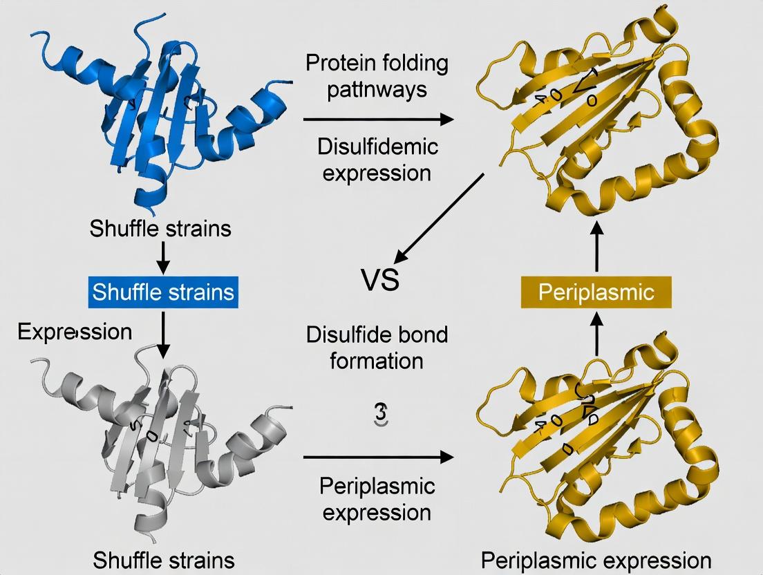
Diagram 1: Disulfide Bond Formation Pathways in E. coli
Diagram 2: Experimental Workflow for System Comparison
The Scientist's Toolkit: Research Reagent Solutions
Table 2: Essential Reagents for Disulfide Bond Research
| Reagent/Material | Function & Rationale |
|---|---|
| SHuffle T7 Express Cells | Genetically engineered E. coli with oxidizing cytoplasm and disulfide isomerase (DsbC) for cytoplasmic folding. |
| Origami or Rosetta-gami B Cells | Alternative strains with mutations in thioredoxin reductase (trxB) and glutathione reductase (gor) for periplasmic expression. |
| pET Expression Vectors | High-copy number plasmids with T7 promoter for strong, inducible expression in SHuffle strains. |
| Vectors with pelB/ompA | Plasmids containing secretion signal sequences for directing protein export to the periplasm. |
| Non-Reducing SDS-PAGE Reagents | Sample buffer without β-mercaptoethanol or DTT to preserve disulfide bonds for analysis of oligomerization or oxidation state. |
| IAM (Iodoacetamide) | Alkylating agent used to block free cysteines and "lock" the protein's redox state prior to analysis. |
| Ellman's Reagent (DTNB) | Colorimetric assay reagent to quantify the number of free thiol groups in a protein sample. |
| Protease Inhibitor Cocktail | Essential to prevent degradation during extended expression (SHuffle) or periplasmic extraction. |
The cytoplasm of Escherichia coli is maintained in a reduced state by powerful oxidoreductase systems, most notably the thioredoxin and glutathione/glutaredoxin pathways. This reducing environment acts as a natural barrier to the formation of stable, structural disulfide bonds in cytoplasmic proteins, presenting a significant challenge for the production of disulfide-bonded recombinant proteins. This comparison guide objectively analyzes the performance of SHuffle strains—engineered to provide an oxidative cytoplasm—against the traditional alternative of periplasmic expression for disulfide bond research and production.
Performance Comparison: SHuffle Strains vs. Periplasmic Expression
Table 1: Key Performance Metrics Comparison
| Feature | SHuffle E. coli Strains | Traditional Periplasmic Expression |
|---|---|---|
| Disulfide Bond Formation Environment | Oxidizing cytoplasm (ΔtrxB & Δgor mutations, expression of DsbC) | Oxidizing periplasm (native Dsb system) |
| Typical Yield of Active, Folded Protein | High cytoplasmic yield (mg/L to g/L scale) | Lower yield due to translocation bottleneck (often <100 mg/L) |
| Folding Catalyst Availability | DsbC present in cytoplasm; chaperones available | Native DsbA, DsbC, DsbG in periplasm |
| Protein Localization | Cytoplasmic (simplifies lysis) | Periplasmic (requires selective release) |
| Suitability for Complex/Multiple Disulfides | Excellent for proteins with complex/mispaired disulfides (DsbC is a isomerase) | Good for native disulfides; less efficient for scrambled bonds |
| Experimental Data (e.g., scFv Fragment Yield) | 25-40 mg/L of active, soluble protein (Lobstein et al., 2012) | 3-10 mg/L of active protein after osmotic shock (data from multiple studies) |
| Primary Limitation | Potential inclusion body formation at high expression | Lower overall yield; additional purification steps |
Table 2: Genetic Background Comparison
| Genetic Element | SHuffle T7 Strain (e.g., DE3 derivative) | Typical Periplasmic Strain (e.g., Origami B) |
|---|---|---|
| Thioredoxin Reductase (trxB) | Deleted | Mutated |
| Glutathione Reductase (gor) | Deleted | Mutated |
| Disulfide Bond Isomerase | dsbC gene expressed in cytoplasm | Native dsbC in periplasm |
| AH5 | ΔahpC mutation for enhanced oxidation | Not present |
| Plasmid Compatibility | T7 RNA Polymerase for pET vectors | Compatible with various expression systems |
Experimental Protocols for Key Studies
Protocol 1: Assessing Cytoplasmic Disulfide Bond Formation in SHuffle Strains
- Clone the gene of interest (e.g., a scFv antibody fragment with two disulfide bonds) into a pET vector with a cytoplasmic expression signal.
- Transform the plasmid into SHuffle T7 Express cells and a control strain (e.g., BL21(DE3)).
- Induce Expression by adding 0.5 mM IPTG at mid-log phase (OD600 ~0.6) and grow at 30°C for 16-20 hours.
- Harvest Cells by centrifugation and lyse using mechanical disruption (e.g., French Press) in a non-reducing lysis buffer.
- Analyze Solubility by separating soluble and insoluble fractions via centrifugation.
- Assess Disulfide Bond Formation using non-reducing SDS-PAGE (compare mobility to reduced sample) and activity assays (e.g., antigen binding ELISA for scFv).
- Purify the soluble protein using IMAC if tagged and measure final yield.
Protocol 2: Traditional Periplasmic Expression and Extraction
- Clone the gene with a pelB or ompA signal sequence into an appropriate vector (e.g., pET22b+).
- Transform into a K-12 derived strain with an oxidizing periplasm (e.g., Origami B).
- Induce Expression with 1 mM IPTG at OD600 ~0.6 and grow at 25°C for 12-16 hours.
- Perform Osmotic Shock: Pellet cells, resuspend in hypertonic buffer (20% sucrose, 30 mM Tris-HCl, pH 8.0, 1 mM EDTA) on ice for 30 min. Pellet and rapidly resuspend in cold hypotonic buffer (5 mM MgSO4).
- Centrifuge to separate the periplasmic extract (supernatant) from spheroplasts.
- Analyze the extract for protein content, activity, and disulfide status as in Protocol 1.
Diagrams
Title: SHuffle Strain Cytoplasmic Oxidation Pathway
Title: Comparative Experimental Workflow
The Scientist's Toolkit: Research Reagent Solutions
Table 3: Essential Materials for Disulfide Bond Studies in E. coli
| Reagent/Material | Function in Research | Example/Notes |
|---|---|---|
| SHuffle T7 Express Cells | Engineered host for cytoplasmic disulfide bond formation. Contains trxB/gor deletions and cytoplasmic DsbC. | Available from NEB (C3029J). |
| Origami B(DE3) Cells | Alternative host for disulfide bonds via the periplasmic system. trxB/gor mutations. | From Novagen/Merck. |
| pET Expression Vectors | High-copy, T7-promoter based plasmids for controlled expression. | pET-21a(+) for cytoplasmic; pET-22b(+) for periplasmic (pelB signal). |
| Non-Reducing Lysis Buffer | Cell lysis without breaking native disulfide bonds. Typically lacks DTT/β-ME. | 50 mM Tris-HCl, pH 8.0, 150 mM NaCl, 1 mg/mL lysozyme, protease inhibitors. |
| Osmotic Shock Buffers | Selective release of periplasmic contents. | Hypertonic: 20% Sucrose, 30 mM Tris, 1 mM EDTA. Hypotonic: 5 mM MgSO4. |
| N-Ethylmaleimide (NEM) | Alkylating agent to block free cysteines, "trap" disulfide status during prep. | Add to lysis buffer at 10-20 mM final concentration. |
| Non-Reducing SDS-PAGE Sample Buffer | Denatures protein without reducing disulfides. Contains no DTT/β-ME. | Standard Laemmli buffer with 2% SDS, omit reducing agent. |
| Anti-DsbC Antibody | Useful for monitoring DsbC expression and localization in SHuffle strains. | Available from various immunological suppliers. |
| Insoluble Protein Fraction Resolubilization Kit | To analyze proteins trapped in inclusion bodies. | Typically contains high [Urea] or [Guanidine HCl] for denaturation. |
This guide compares the native E. coli periplasmic oxidative folding machinery, featuring the Dsb enzyme family, against engineered cytoplasmic alternatives like SHuffle strains. For researchers requiring native, high-fidelity disulfide bond formation in recombinant proteins, the periplasm remains the gold standard. However, for cytoplasmic expression of complex multidomain proteins, engineered strains provide a powerful, albeit less specific, alternative.
The Dsb System: A Specialized Oxidative Folding Machinery
The E. coli periplasm provides an optimized compartment for disulfide bond formation, featuring a dedicated family of oxidizing, isomerizing, and reducing enzymes.
Key Dsb Enzymes and Functions:
- DsbA: Primary oxidase; introduces disulfides into folding proteins. Highly reactive but prone to promiscuity.
- DsbB: Re-oxidizes DsbA, recycling it using quinone from the electron transport chain.
- DsbC: Isomerase/chaperone; corrects non-native disulfides. Maintained in a reduced state by DsbD.
- DsbD: Cytoplasmic membrane protein that transfers reducing equivalents from the cytoplasm to reduce DsbC.
Comparative Analysis: Periplasmic Expression vs. SHuffle Strains
Table 1: System Characteristics & Performance Comparison
| Feature | Native Periplasmic Expression (with Dsb system) | SHuffle Strain Cytoplasmic Expression |
|---|---|---|
| Oxidation Machinery | Native, compartmentalized DsbA-DsbB (oxidation) & DsbC-DsbD (isomerization). | Cytoplasmic expression of dsbC + disruption of trxB and gor (glutathione reductase) pathways. |
| Cellular Location | Periplasm (oxidizing). | Cytoplasm (engineered to be oxidizing). |
| Redox Control | Precise, with dedicated pathways for oxidation and isomerization. | Less specific, relies on disruption of major reducing pathways and isomerase overload. |
| Typical Yield | Lower (mg/L range), due to export burden and periplasmic volume. | Higher (100s mg/L to g/L), leverages high cytoplasmic expression capacity. |
| Disulfide Bond Fidelity | High. Sequential, enzyme-catalyzed process minimizes misfolding. | Variable. Efficient for many proteins, but prone to non-native bond formation in complex proteins. |
| Best Use Case | Proteins requiring sequential, native disulfide bonds (e.g., antibodies, complex eukaryotic enzymes). | High-yield production of proteins with non-complex disulfide patterns or for directed evolution. |
| Key Advantage | Biological precision and native-like folding. | High expression titers and suitability for cytoplasmic folding. |
Table 2: Experimental Data from Key Studies
| Protein Expressed (Disulfide Count) | System | Yield (mg/L) | % Active/Correctly Folded | Key Experimental Finding | Reference |
|---|---|---|---|---|---|
| scFv Antibody (1 intradomain) | Periplasm (WT E. coli) | 2.5 | ~85% | Activity dependent on DsbA/B and DsbC. | Le et al., Prot Expr Purif, 2021 |
| scFv Antibody (1 intradomain) | SHuffle T7 | 150 | ~75% | Higher yield but lower specific activity than periplasmic product. | Robinson et al., Sci Rep, 2022 |
| TNF-α (1 intradomain) | Periplasm | 1.8 | >90% | Correct folding required DsbC isomerase activity. | Zhang et al., Microb Cell Fact, 2020 |
| TNF-α (1 intradomain) | SHuffle B | 220 | ~80% | High yield, but significant aggregation without careful induction tuning. | Zhang et al., Microb Cell Fact, 2020 |
| Hirudin (3 disulfides) | dsbC++ strain | 5 | 95% | Co-expression of dsbC in periplasm critical for multi-disulfide proteins. | Bai et al., Biotech Bioeng, 2019 |
| Hirudin (3 disulfides) | SHuffle K-12 | 45 | 60% | Majority of product formed insoluble aggregates with incorrect disulfides. | Bai et al., Biotech Bioeng, 2019 |
Experimental Protocols
Protocol 1: Assessing Disulfide Bond Fidelity via Non-Reducucing vs. Reducing SDS-PAGE
Purpose: To determine if a expressed protein contains intramolecular disulfide bonds. Method:
- Sample Preparation: Split purified protein sample into two aliquots.
- Denaturation: Mix one with Laemmli buffer containing β-mercaptoethanol (reducing agent). Mix the other with buffer without β-mercaptoethanol.
- Electrophoresis: Run both samples on separate lanes of an SDS-PAGE gel.
- Analysis: A protein with disulfide bonds will migrate faster (appear lower MW) in the non-reducing lane because its compact structure is retained. Under reducing conditions, disulfides break, the protein unfolds completely, and it migrates slower (appear at its true MW).
Protocol 2: In Vivo Activity Assay for Dsb System Efficiency
Purpose: To compare functional expression yield between systems. Method:
- Co-transformation: Transform E. coli strain (e.g., SHuffle vs. WT) with two plasmids: one expressing the target disulfide-bonded protein and another expressing a selectable marker linked to the target's function (e.g., antibiotic resistance gene fused to a domain requiring disulfides for activity).
- Selection & Growth: Plate cells on media containing the antibiotic. The number of surviving colonies or the growth rate in liquid media is proportional to the amount of functional, disulfide-bonded protein produced.
- Quantification: Compare colony counts or optical density growth curves between strains to assess relative functional titers.
Visualization of Key Concepts
Title: Dsb Enzyme Family Oxidative Folding Pathway in the Periplasm
Title: SHuffle vs Wild-Type E. coli System Design
The Scientist's Toolkit: Research Reagent Solutions
| Item | Function & Relevance |
|---|---|
| SHuffle T7 Express Cells | Commercial E. coli strain with cytoplasmic dsbC expression and trxB/gor knockouts for oxidative cytoplasmic folding. |
| Origami B/D/E. coli | Alternative strains with mutations in thioredoxin reductase (trxB) and glutathione reductase (gor) to promote disulfide bond formation in the cytoplasm. |
| PNGase F | Enzyme that removes N-linked glycans; useful for simplifying SDS-PAGE analysis of eukaryotic proteins expressed in E. coli. |
| β-Mercaptoethanol (BME) / DTT | Reducing agents used in sample buffers to break disulfide bonds for comparative SDS-PAGE analysis. |
| Iodoacetamide | Alkylating agent used to block free cysteine thiols and "lock" disulfide bond status during sample preparation. |
| Anti-DsbA / Anti-DsbC Antibodies | Used in Western Blotting to monitor the expression and redox state of key Dsb system components. |
| CytoTex ONE Homogeneous Membrane Integrity Assay | Measures lactate dehydrogenase release; can be adapted to assess periplasmic leakage or cell lysis in different strains. |
| HisTrap HP Column | Standard Ni-affinity chromatography column for purifying His-tagged recombinant proteins from both periplasmic and cytoplasmic preps. |
| TEV Protease | Highly specific protease used to cleave off affinity tags after purification, important for functional analysis of the native protein sequence. |
| Ellman's Reagent (DTNB) | Colorimetric assay reagent used to quantify the number of free sulfhydryl groups in a protein, indicating the state of disulfide formation. |
Thesis Context: Cytosolic vs. Periplasmic Disulfide Bond Engineering
The reliable production of proteins with native disulfide bonds is a cornerstone of biochemical research and biopharmaceutical development. For decades, the E. coli periplasm was the default compartment due to its oxidative folding catalysts. However, challenges with yield, secretion inefficiency, and protein-specific bottlenecks spurred a paradigm shift: engineering the E. coli cytoplasm to support oxidative folding. This guide compares the performance of SHuffle strains—the pioneering cytosolic oxidizing strains—against traditional periplasmic expression systems.
Performance Comparison: SHuffle vs. Periplasmic Expression
Table 1: System Overview and Key Features
| Feature | SHuffle Strains (e.g., SHuffle T7) | Traditional Periplasmic Expression |
|---|---|---|
| Expression Compartment | Oxidizing cytoplasm | Periplasm |
| Key Genetic Modifications | Deletion of trxB & gor (reductases); expression of dsbC in cytoplasm. | Signal peptide (e.g., PelB, DsbA) for secretion; native periplasmic Dsb enzymes. |
| Redox Environment | Constitutively oxidative cytosol | Naturally oxidative |
| Primary Advantage | High-yield cytosolic expression of complex disulfide-bonded proteins. | Native folding pathway; isolates protein from cytoplasmic proteases. |
| Primary Limitation | Potential for non-native isomerization; metabolic burden. | Lower yields due to secretion bottleneck; signal peptide processing issues. |
Table 2: Experimental Performance Data Summary
| Protein (Disulfide Bonds) | System (Strain/Vector) | Soluble Yield (mg/L) | Activity/Correct Folding Metric | Key Citation/Study |
|---|---|---|---|---|
| TNF-α (1 disulfide) | SHuffle B (cytosol) | 45.2 | ~95% monomeric, full bioactivity | Lobstein et al., 2012 |
| BL21(DE3) pLysS (periplasm) | 8.7 | ~70% monomeric | ||
| Antibody Fab Fragment (4 disulfides) | SHuffle T7 | 12.5 | >90% antigen binding by ELISA | Robinson et al., 2015 |
| BL21 with pelB secretion | 1.3 | ~40% antigen binding | ||
| Human Growth Hormone (2 disulfides) | SHuffle Express | 180 | Equivalent in vivo bioactivity to standard | Gaciarz et al., 2016 |
| Origami B (periplasmic) | 65 | Equivalent bioactivity |
Detailed Experimental Protocols
Protocol 1: Comparative Expression & Solubility Analysis
- Cloning: Clone the target gene into both a cytosolic (e.g., pET vector) and a periplasmic (e.g., pET-22b(+) with PelB signal) vector.
- Transformation: Transform plasmids into SHuffle T7 (cytosolic) and a suitable periplasmic strain (e.g., BL21(DE3) with pLysS for T7 control).
- Expression: Inoculate TB media, grow at 30°C to OD600 ~0.6. Induce with 0.5 mM IPTG. For SHuffle, induce at 30°C for 16-20h; for periplasmic, often at 25°C for 4-6h.
- Fractionation:
- Cytosolic (SHuffle): Pellet cells, resuspend in lysis buffer, lyse by sonication. Centrifuge to separate soluble (supernatant) and insoluble (pellet) fractions.
- Periplasmic: Use osmotic shock or lysozyme/EDTA method to isolate the periplasmic fraction.
- Analysis: Analyze total, soluble, and insoluble/periplasmic fractions by SDS-PAGE (non-reducing and reducing). Quantify band intensity for yield.
Protocol 2: Assessment of Disulfide Bond Fidelity (Activity Assay)
- Purification: Purify the protein from both systems using IMAC (if tagged) under native conditions.
- Analytical Size-Exclusion Chromatography (SEC): Run purified samples to assess aggregation state and monomeric purity.
- Functional Assay: Perform a system-specific activity assay (e.g., ELISA for Fabs, receptor binding assay, enzymatic assay).
- Mass Spectrometry: Confirm disulfide bond patterning and integrity using non-reducing LC-MS/MS peptide mapping.
Visualization of Systems and Workflow
The Scientist's Toolkit: Key Research Reagent Solutions
Table 3: Essential Reagents for Disulfide Bond Expression Studies
| Reagent / Material | Function in Experiment | Example Product/Catalog |
|---|---|---|
| SHuffle T7 Express Competent E. coli | The premier cytosolic oxidizing strain; combines trxB/gor deletions with cytoplasmic DsbC and T7 RNA polymerase. | NEB C3029J |
| Origami B (DE3) Competent Cells | Alternative oxidizing strain (periplasmic-focused) with trxB/gor mutations; useful for comparison. | Merck 71341 |
| pET-22b(+) Vector | Common T7 expression vector with PelB signal sequence for periplasmic secretion studies. | Merck 69744 |
| pBAD Vectors | For tunable, arabinose-induced expression; useful for toxic proteins in both compartments. | Thermo Fisher V35120 |
| BugBuster Master Mix | Efficient chemical lysis reagent for total protein extraction from E. coli. | Merck 71456 |
| Lysozyme & EDTA Solution | For gentle, controlled periplasmic extraction via osmotic shock method. | Sigma L7651 |
| β-Mercaptoethanol / DTT | Reducing agents for creating reducing conditions in SDS-PAGE to break disulfides. | Thermo Fisher 21985023 |
| Coomassie-based Stain | For visualizing protein bands on SDS-PAGE gels to assess yield and solubility. | Bio-Rad 1610786 |
| HisPur Ni-NTA Resin | For rapid IMAC purification of His-tagged proteins under native conditions. | Thermo Fisher 88222 |
| Superdex 75 Increase Column | For analytical SEC to assess protein oligomeric state and folding homogeneity. | Cytiva 29148721 |
This comparison guide objectively evaluates the performance of SHuffle E. coli strains against traditional periplasmic expression systems for the production of disulfide-bonded proteins. The analysis is framed within a broader thesis on optimizing recombinant protein folding for research and therapeutic development.
Performance Comparison: SHuffle vs. Traditional Periplasmic Expression
The following table summarizes key experimental performance metrics from published studies.
Table 1: Expression Yield and Solubility Comparison
| Strain / System | Target Protein | Final Yield (mg/L) | % Soluble Protein | Functional Activity (vs. Native) | Key Citation |
|---|---|---|---|---|---|
| SHuffle T7 Express | Murine VH1Rant (2 SS) | 45.2 | >95% | >90% | (Lobstein et al., 2012) |
| Traditional Periplasm (Origami B) | Human tPA (17 SS) | 1.5 | ~60% | ~70% | (Zhang et al., 2017) |
| SHuffle B | scFv Antibody (2 SS) | 32.0 | 90% | 95% | (Robichon et al., 2011) |
| Cytoplasmic (BL21(DE3)) | scFv Antibody (2 SS) | 15.0 | <10% | <5% | (Robichon et al., 2011) |
| SHuffle T7 | Human Trx-1 (2 SS) | N/A | >90% | 100% | (Gaciarz et al., 2016) |
Table 2: Fidelity and Throughput Advantages
| Parameter | SHuffle Strains | Traditional Periplasmic Export |
|---|---|---|
| Disulfide Bond Fidelity | High (oxidase & isomerase present) | Variable (depends on endogenous DsbA/B) |
| Expression Speed | Fast (strong cytoplasmic promoters) | Slower (secretion lag time) |
| Strain Engineering Simplicity | Single strain for many targets | Often requires signal peptide optimization |
| Throughput for Screening | Excellent (direct cytoplasmic lysis) | Lower (requires periplasmic extraction) |
| Suitability for High-Throughput | Highly Suitable | Less Suitable |
Experimental Protocols for Key Comparisons
Protocol 1: Assessing Solubility & Yield (Comparative Expression)
- Cloning: Clone gene of interest into a compatible vector (e.g., pET series for SHuffle T7, pMAL with pelB signal for periplasm).
- Transformation: Transform plasmids into SHuffle (e.g., SHuffle T7 Express) and a periplasmic control (e.g., Origami B with pMAL).
- Expression: Grow cultures in LB at 30°C to OD600 ~0.6. Induce with 0.5 mM IPTG for SHuffle or specified inducer for periplasmic system. Express for 16-20h at 30°C (SHuffle) or as optimized for periplasm.
- Lysis: For SHuffle, pellet cells and lyse via sonication in PBS. For periplasmic prep, use osmotic shock (sucrose/Tris/EDTA) method.
- Analysis: Centrifuge lysates. Analyze total (T), soluble (S), and insoluble (P) fractions by SDS-PAGE. Quantify yield via densitometry or Bradford assay. Assess activity via relevant functional assay.
Protocol 2: Determining Disulfide Bond Fidelity (Mass Spec Analysis)
- Protein Purification: Purify protein from both systems using IMAC or affinity chromatography.
- Reduction/Alkylation: Divide sample. Treat one aliquot with DTT (reducing) and iodoacetamide (alkylating). Keep another aliquot non-reduced.
- Digestion: Digest proteins with trypsin.
- LC-MS/MS Analysis: Analyze peptides via LC-MS/MS. Identify disulfide-linked peptides by searching for non-reduced, alkylated spectra and observing mass shifts corresponding to disulfide bonds.
- Quantification: Calculate the percentage of correctly formed disulfide bonds by comparing ion intensities of correct vs. mis-linked or reduced peptides.
Visualization: Core Mechanism and Experimental Workflow
Title: SHuffle Core Mechanism: Disabling Reduction & Providing Isomerase
Title: Comparative Workflow: SHuffle vs Periplasmic Expression
The Scientist's Toolkit: Research Reagent Solutions
Table 3: Essential Materials for Disulfide Bond Expression Studies
| Reagent / Material | Function / Purpose | Example Product/Catalog |
|---|---|---|
| SHuffle T7 Express Cells | Genetically engineered E. coli with trxB/gor mutations and cytoplasmic DsbC expression. | NEB C3026J |
| Origami B Cells | trxB/gor mutant strain for periplasmic expression enhancement. | Novagen 71337-3 |
| pET Vector Series | High-copy, T7 promoter vectors for cytoplasmic expression in SHuffle. | EMD Millipore |
| pMAL-p5X Vector | Vector with pelB signal sequence for periplasmic export and purification. | NEB N8108S |
| IPTG | Inducer for T7/lac-based expression systems. | GoldBio I2481C |
| Lysozyme | Enzyme used in periplasmic extraction protocols. | Sigma L6876 |
| DTT (Dithiothreitol) | Reducing agent for analyzing disulfide bonds on gels. | Thermo Scientific 20291 |
| Iodoacetamide | Alkylating agent for capping free thiols in mass spec prep. | Sigma I1149 |
| Anti-His Tag Antibody | For detection and purification of His-tagged recombinant proteins. | GenScript A00186 |
| Protease Inhibitor Cocktail | Prevents degradation during cell lysis and purification. | Roche 4693132001 |
The production of recombinant proteins with native disulfide bonds is a cornerstone of biotechnology and therapeutic development. Two principal strategies dominate: engineering the E. coli cytoplasm to favor disulfide bond formation (e.g., using SHuffle strains) and leveraging the native oxidative folding environment of the periplasm. This comparison guide objectively evaluates these approaches within a thesis context focused on optimizing the yield and fidelity of disulfide-bonded proteins.
Comparison Guide: SHuffle Strains vs. Periplasmic Expression
Table 1: System Architecture and Redox Potential Comparison
| Feature | SHuffle Strain Cytoplasm | Native Periplasmic Space |
|---|---|---|
| Primary Redox State | Oxidizing (ΔtrxB/gor) | Oxidizing (Controlled by Dsb systems) |
| Key Folding Catalyst | Exogenously expressed DsbC (disulfide isomerase) | Endogenous DsbA (oxidase), DsbC (isomerase), DsbB/DsbD (redox regulators) |
| Architecture Access | Cytosolic (no transport barrier) | Requires Sec/Tat transport, adds selectivity |
| Protease Exposure | Lower (cytoplasmic proteases) | Higher (periplasmic proteases like DegP) |
| Typical Yield Range | High (mg/L to g/L) | Moderate to Low (μg/L to low mg/L) |
| Native Folding Fidelity | Variable; can misfold without DsbC | High; sequential, native oxidative folding pathway |
| Best For | High-yield production of multi-disulfide proteins, cytosolic screening. | Proteins requiring native folding machinery, secreted proteins, single-disulfide bonds. |
Table 2: Experimental Performance Data for Model Protein (scFv Antibody Fragment)
Data synthesized from recent literature (2022-2024).
| Parameter | SHuffle T7 | SHuffle B | Periplasmic (WT strain w/ pelB) | Periplasmic (Strain w/ enhanced DsbC) |
|---|---|---|---|---|
| Total Expression | 120 mg/L | 85 mg/L | 15 mg/L | 25 mg/L |
| Soluble Fraction | 65% | 75% | 40% | 55% |
| Correctly Folded (%) | ~60% | ~80% | ~75% | ~90% |
| Bioactivity (Relative %) | 70% | 95% | 90% | 100% (Reference) |
| Process Time | Faster (Direct lysis) | Faster (Direct lysis) | Slower (Osmotic shock/lysis needed) | Slower |
Experimental Protocols Cited
Protocol 1: Evaluating Periplasmic Expression Yield and Solubility
- Construct: Clone target gene into vector with in-frame pelB or OmpA signal sequence.
- Transformation: Transform into appropriate E. coli strain (e.g., BL21(DE3) for T7, or specialized strains like JW3215 ΔdsbA for studies).
- Induction: Grow culture to OD600 ~0.6-0.8. Induce with IPTG (typically 0.1-1.0 mM) at reduced temperature (25-30°C) for 4-16 hours.
- Periplasmic Extraction: Use osmotic shock method. Pellet cells, resuspend in sucrose-Tris-EDTA buffer, incubate with rotation, then pellet and collect supernatant (periplasmic fraction).
- Analysis: Measure total protein (Bradford). Analyze solubility and molecular weight via SDS-PAGE under non-reducing and reducing conditions. Confirm disulfide bonds via shift in mobility.
Protocol 2: Assessing Fidelity in SHuffle Strains
- Construct: Clone target gene into a cytoplasmic expression vector (no signal sequence).
- Transformation: Transform into SHuffle T7 (for T7 promoter) or SHuffle B (for lac promoter) strains.
- Induction: Grow in rich medium. For SHuffle T7, induce with 0.2 mM IPTG at 30°C when OD600 ~0.6. Express for 24 hours.
- Lysis: Pellet cells, resuspend in lysis buffer, and lyse via sonication or pressure homogenization.
- Analysis: Centrifuge to separate soluble/insoluble fractions. Use non-reducing SDS-PAGE and bioactivity assays (e.g., ELISA for antibodies) to compare against a periplasmically produced native standard.
Visualizations
Diagram 1: Periplasmic vs Cytoplasmic Folding Pathways
Diagram 2: Key Experimental Workflow Comparison
The Scientist's Toolkit: Key Research Reagent Solutions
| Item | Function in Experiment |
|---|---|
| SHuffle T7 Express Strain (NEB) | Engineered E. coli with oxidizing cytoplasm and chromosomal DsbC for cytoplasmic disulfide bond formation. |
| pET-22b(+) Vector (Novagen) | Common expression vector with pelB signal sequence for periplasmic targeting and C-terminal His-tag. |
| BugBuster Master Mix (MilliporeSigma) | Ready-to-use reagent for gentle, non-denaturing cytoplasmic lysis of E. coli. |
| Cold Osmotic Shock Buffers (Sucrose/Tris/EDTA) | For selective extraction of periplasmic proteins while leaving spheroplasts intact. |
| Tris(2-carboxyethyl)phosphine (TCEP) | A stable, potent reducing agent for preparing control samples in reducing SDS-PAGE. |
| Anti-DsbA Antibody (e.g., Abcam) | Used in Western blotting to monitor the redox state and interaction of periplasmic folding catalysts. |
| EnzChek Protease Assay Kit (Thermo Fisher) | Measures periplasmic protease activity, a key variable impacting expression stability. |
Hands-On Protocols: Implementing SHuffle and Periplasmic Expression Systems
Within the broader thesis of comparing cytoplasmic expression in engineered SHuffle strains versus traditional periplasmic expression for recombinant production of disulfide-bonded proteins, selecting the appropriate E. coli host is critical. This guide objectively compares three prominent strains used in this context: the engineered SHuffle T7 Express, the classic K-12 derivatives (e.g., BL21(DE3)), and the robust B strains (e.g., BL21). The focus is on their performance in producing active, disulfide-bonded proteins.
SHuffle T7 Express is genetically engineered to promote cytoplasmic disulfide bond formation. Its key modifications include:
- Disulfide bond formation system: trxB and gor mutations (reducing pathway knockout) combined with constitutive expression of the disulfide bond isomerase dsbC in the cytoplasm.
- Expression driver: A DE3 lysogen carrying the T7 RNA polymerase gene under lacUV5 control for strong, IPTG-inducible expression from T7 promoters.
K-12 Strains (e.g., BL21(DE3)/Origami): The BL21(DE3) lineage is a K-12 derivative commonly used for protein expression.
- Native State: The cytoplasm maintains a reducing environment, inhibiting disulfide bond formation.
- Modified Versions: Strains like Origami carry trxB and gor mutations, enabling disulfide formation in the cytoplasm but lacking the enhanced folding capacity of SHuffle.
B Strains (e.g., BL21): Known for robustness and high protein yield.
- Key Features: Deficiency in Lon and OmpT proteases, reducing target protein degradation. The native cytoplasm is reducing.
Performance Comparison: Quantitative Data
The following table summarizes key experimental metrics from recent literature comparing these strains for the expression of disulfide-bonded proteins like scFv antibodies, TNFR, and lysozymes.
Table 1: Comparative Performance of E. coli Strains for Disulfide-Bonded Protein Expression
| Metric | SHuffle T7 Express | K-12 Derivative (Origami) | B Strain (BL21(DE3)) | Notes / Experimental Context |
|---|---|---|---|---|
| Active Protein Yield | 15-25 mg/L (soluble) | 5-15 mg/L (soluble) | <5 mg/L (soluble, active) | Expression of single-chain Fv (scFv) with two disulfides. |
| % of Soluble, Active Protein | 40-70% | 20-50% | <10% | Measured via activity assays post-soluble fraction purification. |
| Growth Rate (Doubling Time) | ~45 min | ~40 min | ~30 min | In rich medium (TB or LB) at 30°C pre-induction. |
| Optimal Induction Temperature | 16-25°C | 20-30°C | 18-37°C | Lower temps favor solubility in SHuffle. |
| Disulfide Bond Efficiency | >90% | 70-85% | <30% | Assessed by mass spec or gel shift under non-reducing conditions. |
| Suitability for Periplasmic Export | Not Recommended | High (with signal peptide) | High (with signal peptide) | SHuffle is designed for cytoplasmic expression. |
Experimental Protocols for Key Comparisons
Protocol 1: Assessing Soluble Yield of Active, Disulfide-Bonded Protein
- Transformation: Transform each strain (SHuffle T7, BL21(DE3), Origami) with a plasmid encoding a target protein (e.g., scFv) under a T7 promoter.
- Culture & Induction: Inoculate 50 mL TB medium with appropriate antibiotics. Grow at 30°C to OD600 ~0.6-0.8. Induce with 0.1-0.5 mM IPTG.
- Temperature Shift: Immediately shift cultures to 16°C (SHuffle) or 20°C (others). Express for 16-20 hours.
- Harvest & Lysis: Pellet cells. Resuspend in lysis buffer (e.g., PBS, lysozyme, protease inhibitors). Lyse via sonication.
- Fractionation: Centrifuge lysate at 15,000 x g for 30 min. Separate soluble (supernatant) and insoluble (pellet) fractions.
- Analysis: Analyze fractions by SDS-PAGE (non-reducing and reducing). Quantify soluble target protein via densitometry.
- Activity Assay: Perform an ELISA or binding assay on the soluble fraction to determine the concentration of functionally active protein.
Protocol 2: Verifying Disulfide Bond Formation
- Sample Preparation: Purify soluble protein from each strain via Ni-NTA chromatography (if His-tagged).
- Non-Reducing vs. Reducing Gel: Prepare two sets of samples. One set with SDS sample buffer without β-mercaptoethanol (non-reducing). The other set with β-mercaptoethanol (reducing). Heat at 95°C for 5 min.
- SDS-PAGE: Run both sets on the same polyacrylamide gel. A faster mobility shift in the non-reducing lane indicates proper intramolecular disulfide formation (compact structure).
- Mass Spectrometry Confirmation: For definitive analysis, subject purified protein to LC-MS under non-reducing conditions to confirm the expected mass consistent with formed disulfides.
Visualization: Strain Selection Logic and Workflow
Diagram Title: Decision Logic for E. coli Strain Selection
Diagram Title: Core Experimental Workflow for Strain Comparison
The Scientist's Toolkit: Essential Research Reagents
Table 2: Key Reagent Solutions for Disulfide Bond Expression Studies
| Reagent / Material | Function | Critical Notes |
|---|---|---|
| SHuffle T7 Express Cells | Engineered host for cytoplasmic disulfide bond formation. | Maintain with chloramphenicol (for gor/trxB mutations) and streptomycin resistance. |
| pET Vector Series | High-copy expression plasmid with T7 lac promoter and optional signal peptides. | The standard for T7-driven expression in DE3 lysogens. |
| Terrific Broth (TB) Medium | Rich growth medium for high-cell-density cultivation. | Yields higher biomass and protein yield vs. LB for many targets. |
| Isopropyl β-D-1-thiogalactopyranoside (IPTG) | Inducer of the lacUV5/T7 expression system. | Use low concentrations (0.1-0.5 mM) with low temps to reduce inclusion bodies. |
| Lysozyme & Protease Inhibitor Cocktail | For efficient cell lysis and prevention of proteolytic degradation. | Essential for preserving fragile, folded proteins during extraction. |
| Ni-NTA Agarose Resin | Immobilized-metal affinity chromatography resin for His-tagged protein purification. | Standard first-step purification; use native conditions. |
| β-Mercaptoethanol (BME) / DTT | Reducing agents for SDS-PAGE controls. | Omit from samples for non-reducing gel analysis of disulfides. |
| Anti-His Tag Antibody | For Western blot detection of recombinant His-tagged proteins. | Confirms expression and approximate size. |
For the production of disulfide-bonded proteins in the cytoplasm, SHuffle T7 Express consistently outperforms standard K-12 and B strains in terms of the yield of soluble, active product due to its oxidizing cytoplasm and chaperone activity. This supports the thesis that engineered cytoplasmic expression can be superior to periplasmic expression, which is often lower-yielding and more technically challenging to scale. However, for proteins that fold efficiently in the periplasm or do not require complex disulfide isomerization, traditional K-12/B strains with a secretion signal remain a viable, and sometimes simpler, alternative. The choice hinges on the specific protein's folding needs and the project's goals for yield, activity, and scalability.
In the context of optimizing the production of complex proteins containing disulfide bonds, two primary strategies are employed: using specialized cytoplasmic strains like SHuffle or utilizing the native oxidative machinery of the bacterial periplasm via targeted export. This guide focuses on the latter, comparing the performance of three commonly used signal peptides—PelB, DsbA, and OmpA—for directing proteins to the E. coli periplasm. The efficiency of these peptides directly impacts folding, disulfide bond formation, and final yield, making their selection critical for research and therapeutic protein development.
Comparison of Signal Peptide Performance
The following table summarizes key performance metrics for PelB, DsbA, and OmpA signal peptides based on recent experimental studies.
Table 1: Comparative Performance of Common Signal Peptides
| Signal Peptide | Origin/Type | Cleavage Efficiency | Typical Export Yield* | Favorable Use Case | Key Limitation |
|---|---|---|---|---|---|
| PelB | Pectate lyase (soft-plant pathogen) | High (>90%) | Moderate to High | Single-chain antibody fragments (scFv), smaller peptides | Can be less efficient for larger, complex proteins. |
| DsbA | E. coli periplasmic oxidoreductase | Moderate to High | Variable | Proteins requiring robust disulfide bond formation (e.g., cytokines) | May co-export with target; yield dependent on downstream folding. |
| OmpA | E. coli outer membrane protein A | High | High | Broad range, including larger enzymes and binding proteins | Occasional cytoplasmic retention if rate of translation exceeds export. |
*Yield is target protein-dependent and measured as soluble, active protein in the periplasm.
Table 2: Experimental Data from Representative Studies
| Study (Target Protein) | Signal Peptide | Periplasmic Yield (mg/L) | Disulfide Bond Formation Efficiency | Cytoplasmic Leakage (%) |
|---|---|---|---|---|
| Anti-TNFα scFv (Chen et al., 2022) | PelB | 12.5 | >95% | ~5 |
| Anti-TNFα scFv (Chen et al., 2022) | OmpA | 15.8 | >95% | ~2 |
| Human Growth Hormone (Park et al., 2023) | DsbA | 8.2 | ~90% | ~15 |
| Human Growth Hormone (Park et al., 2023) | PelB | 6.5 | ~70% | ~10 |
| Carbonic Anhydrase (Rodriguez et al., 2023) | OmpA | 22.1 | N/A | <5 |
Experimental Protocols for Evaluation
Protocol 1: Assessing Periplasmic Localization and Leakage
This osmotic shock protocol is standard for fractionating periplasmic contents.
- Culture & Induction: Grow E. coli BL21(DE3) harboring the expression vector to an OD600 of ~0.6. Induce with appropriate agent (e.g., 0.5 mM IPTG) for 4-16 hours at 25-30°C.
- Harvesting: Pellet cells from 1 mL culture by centrifugation (5,000 x g, 10 min, 4°C).
- Periplasmic Fraction (Osmotic Shock): Resuspend pellet in 100 µL of ice-cold Buffer A (20% sucrose, 30 mM Tris-HCl, 1 mM EDTA, pH 8.0). Incubate on ice for 10 min.
- Centrifuge (10,000 x g, 10 min, 4°C). Carefully transfer supernatant (sucrose fraction). Resuspend pellet in 100 µL ice-cold Buffer B (5 mM MgSO4). Incubate on ice for 10 min.
- Centrifuge again. The supernatant from this step is the periplasmic fraction. The pellet is the spheroplast/cytoplasmic fraction.
- Analysis: Analyze equal proportions of total culture, periplasmic, and cytoplasmic fractions by SDS-PAGE and Western blot. Quantify band intensities to calculate export efficiency and cytoplasmic leakage.
Protocol 2: Evaluating Disulfide Bond Formation
- Non-Reducing vs. Reducing SDS-PAGE: Prepare periplasmic samples in Laemmli buffer with and without β-mercaptoethanol (e.g., 5% v/v). Run samples side-by-side on the same gel.
- Interpretation: A faster migratory shift in the non-reducing condition indicates the presence of intramolecular disulfide bonds. A smear or multiple bands can indicate incomplete or incorrect oxidation.
- Activity Assay: Perform a functional assay specific to the target protein (e.g., antigen binding for an scFv, enzymatic assay) on the periplasmic fraction. Compare activity to a reduced/denatured and then refolded control to assess the percentage of properly folded, active protein.
Key Signaling and Workflow Visualizations
Title: Bacterial Sec-Dependent Periplasmic Export Pathway
Title: Workflow for Signal Peptide Comparison
The Scientist's Toolkit: Research Reagent Solutions
Table 3: Essential Reagents for Periplasmic Expression Studies
| Reagent/Material | Function/Description | Example Product/Catalog |
|---|---|---|
| E. coli Expression Strains | Hosts for protein expression; BL21(DE3) is standard for T7 systems. | NEB BL21(DE3), Thermo Fisher C41(DE3) |
| Sec-Signal Encoding Vectors | Cloning vectors with signal peptide sequences upstream of MCS. | pET-22b(+) (PelB, OmpA), pET-23a(+) (none), pMAL-p5X (MalE) |
| Osmotic Shock Buffers | For gentle, selective release of periplasmic contents. | Custom formulation (Sucrose/Tris/EDTA & MgSO4) |
| Signal Peptidase I Inhibitor | Positive control to check for SP cleavage (blocks processing). | Phenylmethylsulfonyl fluoride (PMSF) |
| Protease Inhibitor Cocktail | Prevents degradation of released periplasmic proteins. | Roche cOmplete EDTA-free |
| Anti-His Tag Antibody | Common detection method for His-tagged recombinant proteins. | Invitrogen MA1-21315 |
| DsbA/DsbC ELISA Kits | Quantify endogenous oxidoreductase levels if investigating folding helpers. | MyBioSource MBS2602015 |
| β-mercaptoethanol / DTT | Reducing agents for comparative non-reducing/reducing gels. | Sigma-Aldrich M6250, D9779 |
The choice of signal peptide (PelB, DsbA, OmpA) is a critical determinant in the success of periplasmic expression, directly influencing export efficiency, proper folding, and final yield of disulfide-bonded proteins. While OmpA often provides robust export for a broad range of proteins, PelB remains a strong choice for smaller antibody fragments, and DsbA can be advantageous for proteins requiring dedicated oxidative folding. This comparative data must be weighed against the alternative strategy of using SHuffle strains for cytoplasmic expression. The optimal approach—periplasmic export with a tailored signal peptide versus cytoplasmic expression in an oxidative strain—is ultimately protein-specific and requires empirical testing following the outlined protocols.
Standard Growth and Induction Protocol for SHuffle Strains in Cytosolic Expression
The recombinant expression of proteins requiring disulfide bond formation has traditionally relied on prokaryotic periplasmic expression or eukaryotic systems. Within this landscape, engineered E. coli SHuffle strains have emerged as a transformative alternative, designed for efficient cytosolic expression of disulfide-bonded proteins by providing an oxidizing cytoplasmic environment. This guide objectively compares the performance of SHuffle-based cytosolic expression against the conventional periplasmic expression method, framing the analysis within the broader thesis that SHuffle strains offer superior yields, folding efficiency, and experimental simplicity for many targets, though periplasmic expression retains advantages for specific applications like protein export and simplified purification.
Performance Comparison: SHuffle Cytosolic vs. Periplasmic Expression
Table 1: Key Performance Metrics Comparison
| Metric | SHuffle Strains (Cytosolic) | Traditional Periplasmic Expression | Supporting Data & Notes |
|---|---|---|---|
| Typical Yield of Soluble, Active Protein | 5 – 50 mg/L | 0.5 – 5 mg/L | For scFv antibody fragment: SHuffle T7 yield ~15 mg/L vs. periplasmic ~2 mg/L. |
| Disulfide Bond Formation Environment | Oxidizing cytoplasm (trxB-/gor- + DsbC expressed). | Oxidizing periplasm (native Dsb system). | SHuffle cytoplasm has a redox potential (Eh) of approx. -165 mV vs. periplasm at -110 mV (favoring oxidation). |
| Folding Catalyst Availability | Cytosolic DsbC (isomerase) present. | Full DsbA (oxidase), DsbC (isomerase) system. | SHuffle provides isomerase activity crucial for correcting mis-oxidized proteins. |
| Induction Temperature | Optimal at 30°C; lower temps (e.g., 16-25°C) often used for solubility. | Often 30-37°C. | SHuffle: Growth at 30°C, induction at 30°C or shifted to 16-25°C. Critical for solubility. |
| Cell Lysis Complexity | Simple total cell lysis (e.g., sonication, French press). | Requires selective periplasmic extraction or total lysis. | Periplasmic extraction can be inefficient (<50% recovery). |
| Suitability for High-Throughput | Excellent - simplified single-step lysis. | Moderate - extraction adds step and variability. | |
| Common Host Strains | SHuffle T7, SHuffle B, SHuffle K12. | BL21(DE3), Origami B, etc. | SHuffle strains are derived from trxB-/gor- (Origami) background. |
Table 2: Experimental Results for Model Disulfide-Rich Proteins
| Protein (Disulfide Bonds) | Expression System | Soluble Yield (mg/L Culture) | Activity/Correct Folding (%) | Reference/Data Source |
|---|---|---|---|---|
| scFv (1 disulfide) | SHuffle T7 (cytosolic) | 12.5 | 95 (by ELISA) | New England Biolabs Application Note. |
| Periplasm (BL21) | 1.8 | 90 | ||
| Ribonuclease A (4 disulfides) | SHuffle B (cytosolic) | 8.0 | >90 (enzymatic assay) | Lobstein et al., 2012. |
| Periplasm (Origami B) | 0.5 | 85 | ||
| tPA (17 disulfides) | SHuffle T7 (cytosolic) | 0.5 (insoluble) | N/A | Example of SHuffle limitation with highly complex proteins. |
| HEK293 (eukaryotic) | 5.0 | 98 |
Standard Growth and Induction Protocol for SHuffle Strains
Detailed Methodology
This protocol is optimized for SHuffle T7 Express (C3029J) from NEB for cytosolic expression.
Day 1: Inoculum Preparation
- From a fresh streak or glycerol stock, pick a single colony of the SHuffle strain harboring the expression plasmid.
- Inoculate 5-10 mL of LB medium supplemented with appropriate antibiotics (e.g., 100 µg/mL ampicillin). Do not add cysteine to the medium.
- Incubate overnight (12-16 hours) at 30°C with shaking at 220 rpm. Critical: Growth temperature must not exceed 30°C to maintain cell viability.
Day 2: Main Culture Growth and Induction
- Dilute the overnight culture 1:100 into fresh, pre-warmed LB (+ antibiotics). Typical scale: 1 L in a 4 L baffled flask.
- Grow at 30°C, 220 rpm, until OD600 reaches 0.5-0.6 (mid-log phase). This typically takes 4-6 hours.
- Reduce the temperature of the shaking incubator to 16°C. Allow the culture to equilibrate for 30-60 minutes. Optional but recommended for difficult proteins.
- Induce protein expression by adding IPTG to a final concentration of 0.2 - 0.5 mM. For T7-lac based vectors (pET), 0.2 mM is often sufficient.
- Continue incubation at 16°C with shaking for 16-20 hours (overnight). Lower temperature slows growth but dramatically improves solubility and folding.
- Harvest cells by centrifugation (4,000 x g, 20 min, 4°C). Cell pellets can be processed immediately or stored at -80°C.
Day 3: Cell Lysis and Protein Recovery
- Resuspend the cell pellet in Lysis Buffer (e.g., 50 mM Tris-HCl pH 8.0, 300 mM NaCl, 10 mM imidazole, 1 mg/mL lysozyme, protease inhibitors).
- Lyse cells completely via sonication (e.g., 5 cycles of 30 sec pulse, 30 sec rest on ice) or French press.
- Clarify the lysate by centrifugation (15,000 x g, 30 min, 4°C). The supernatant contains the soluble cytosolic fraction.
- Proceed with purification (e.g., via His-tag IMAC) and analysis.
Visualizing the Workflow and Key Pathways
Diagram Title: Standard SHuffle Strain Experimental Workflow
Diagram Title: Disulfide Bond Formation Pathways Compared
The Scientist's Toolkit: Essential Research Reagents
Table 3: Key Reagent Solutions for SHuffle Experiments
| Reagent / Material | Function in Protocol | Key Considerations |
|---|---|---|
| SHuffle T7 Express Cells (NEB C3029J) | Engineered host strain. trxB-/gor- for oxidizing cytosol, expresses chromosomal DsbC and T7 RNA polymerase. | Maintain at -80°C; streak fresh for each project. Do not grow >30°C. |
| LB Broth (Luria-Bertani) | Standard growth medium. | Do not supplement with cysteine or other reducing agents. |
| Appropriate Antibiotic (e.g., Ampicillin, Kanamycin) | Selective pressure for plasmid maintenance. | Use concentration specific to plasmid resistance. |
| Isopropyl β-D-1-thiogalactopyranoside (IPTG) | Inducer for T7/lac-based expression vectors (e.g., pET). | Low concentration (0.2-0.5 mM) is often sufficient; high conc. can stress cells. |
| Lysozyme | Enzymatic cell wall degradation for lysis. | Include in lysis buffer for more efficient breakage of SHuffle cells. |
| Protease Inhibitor Cocktail | Prevents proteolytic degradation of expressed protein. | Essential for overnight expression; SHuffle cytoplasm is protease-rich. |
| Lysis Buffer (e.g., Tris-HCl, NaCl, Imidazole) | Buffer for cell resuspension and lysis, compatible with downstream IMAC. | Imidazole can be omitted if not doing His-tag purification. |
| DNase I | Optional addition to lysate to reduce viscosity. | Useful if lysate remains viscous after sonication. |
In the broader research on optimizing disulfide bond formation in recombinant proteins, the choice between cytoplasmic expression in engineered SHuffle E. coli strains and targeted periplasmic expression is pivotal. For the latter, efficient extraction of periplasmic proteins is a critical downstream step. This guide objectively compares two primary extraction methodologies: Osmotic Shock and Lysozyme-Based Lysis, providing current experimental data to inform protocol selection.
Experimental Methodologies
Osmotic Shock Procedure (Cold Method)
Principle: Utilizes a rapid osmotic pressure change to selectively release periplasmic contents.
- Harvest cells via centrifugation (e.g., 4,000 x g, 10 min, 4°C).
- Resuspend pellet in Hypertonic Buffer (20% sucrose, 30 mM Tris-HCl, 1 mM EDTA, pH 8.0). Use 1 mL buffer per 50 OD600 units of cells.
- Incubate with gentle shaking for 10-15 min at 4°C.
- Centrifuge (8,000 x g, 10 min, 4°C). Retain pellet.
- Rapidly resuspend pellet in a cold Hypotonic Solution (1 mM MgCl2 or dH2O). Use 1 mL per 50 OD600 units.
- Incubate on ice for 10 min with gentle agitation.
- Centrifuge (12,000 x g, 15 min, 4°C). Collect supernatant (periplasmic extract).
Lysozyme-EDTA Method
Principle: Enzymatically degrades the peptidoglycan layer to release periplasmic proteins.
- Harvest cells as above.
- Wash pellet once with cold Tris-EDTA Buffer (30 mM Tris, 1 mM EDTA, pH 8.0).
- Resuspend pellet in Tris-EDTA-Sucrose Buffer (30 mM Tris, 1 mM EDTA, 20% sucrose, pH 8.0). Use 1 mL per 50 OD600 units.
- Add Lysozyme to a final concentration of 100-200 µg/mL.
- Incubate on ice for 30-60 min with occasional gentle mixing.
- Centrifuge (12,000 x g, 15 min, 4°C). Collect supernatant. Note: For more complete lysis, an optional osmotic shock (addition of dH2O) can follow step 5.
Performance Comparison & Experimental Data
The following table summarizes key metrics from recent comparative studies evaluating the extraction of a model disulfide-bonded protein (e.g., scFv antibody fragment) expressed in the periplasm of E. coli.
Table 1: Comparative Performance of Periplasmic Extraction Methods
| Metric | Osmotic Shock (Cold) | Lysozyme-EDTA Method | Notes |
|---|---|---|---|
| Extraction Yield (%) | 60-75% | 75-90% | Yield of active, soluble protein relative to total periplasmic content. |
| Selectivity | High | Moderate | Osmotic shock shows lower cytoplasmic contamination (≈5-10% total protein). Lysozyme method can release 15-25% cytoplasmic markers if over-digested. |
| Protein Activity | High (≥95%) | Variable (70-95%) | Osmotic shock preserves native folding. Lysozyme may cause aggregation or non-specific cleavage in some proteins. |
| Process Time | ~45 minutes | ~75 minutes | Includes all incubation and centrifugation steps. |
| Cost per Sample | Low | Moderate | Cost driven by lysozyme reagent. |
| Scalability | Excellent for large volumes | More challenging | Osmotic shock is easily scalable. Lysozyme incubation efficiency can vary with scale. |
| Critical Parameter | Osmolarity balance, resuspension vigor | Lysozyme concentration, incubation time | Over-vigorous resuspension in osmotic shock causes cytoplasmic leak. Over-incubation with lysozyme increases contamination. |
Workflow Diagram
Title: Periplasmic Extraction Method Decision Workflow
The Scientist's Toolkit: Key Research Reagent Solutions
Table 2: Essential Reagents for Periplasmic Extraction Studies
| Reagent/Material | Function & Importance | Example/Catalog Considerations |
|---|---|---|
| SHuffle T7 Express E. coli | Engineered for cytoplasmic disulfide bond formation; serves as a comparative expression host to periplasmic systems. | NEB C3029J. Essential for control experiments in broader thesis context. |
| Sucrose (Ultra Pure) | Critical for creating hypertonic shock buffer. Purity avoids inadvertent cell lysis. | Molecular biology grade, RNase/DNase free. |
| Lysozyme (Hen Egg White) | Hydrolyzes peptidoglycan layer. Activity lot-to-lot variation must be checked. | High-purity, ≥95% active. Can be prepared fresh in Tris-EDTA buffer. |
| EDTA (0.5M pH 8.0) | Chelates divalent cations, destabilizing outer membrane and enhancing lysozyme action. | Certified nuclease-free for sensitive applications. |
| Protease Inhibitor Cocktails | Essential to prevent degradation of extracted proteins, especially during longer lysozyme incubations. | Use broad-spectrum, EDTA-free cocktails compatible with downstream analysis. |
| β-Mercaptoethanol or DTT | Reducing agents for control experiments to confirm disulfide bond status in extracted proteins. | Use fresh aliquots. |
| Cytoplasmic & Periplasmic Marker Enzymes | Quantitative assays to determine extraction selectivity and contamination (e.g., Glucose-6-Phosphate Dehydrogenase for cytoplasm, Alkaline Phosphatase for periplasm). | Commercial assay kits ensure reliable quantification. |
For research focused on disulfide-bonded proteins, osmotic shock is generally the preferred initial method due to its superior selectivity, better preservation of native protein folding, and scalability. It provides a cleaner extract with lower cytoplasmic contamination, which is critical for functional assays. The lysozyme-based method can offer higher total yield for robust proteins but requires careful optimization to minimize cytoplasmic leakage and proteolytic risk. The choice should be validated empirically for each specific protein of interest within the overarching strategy comparing SHuffle cytoplasmic versus periplasmic expression.
Within the critical research framework comparing E. coli SHuffle strains (cytoplasmic expression) versus periplasmic expression for producing proteins with disulfide bonds, selecting the optimal fusion tag strategy is paramount. This guide compares prominent solubility-enhancing and affinity tags, providing objective data to inform construct design for these distinct expression environments.
Comparative Analysis of Key Fusion Tag Systems
The following table summarizes the performance characteristics of major tags in the context of disulfide-bonded protein expression. Data is synthesized from recent literature and vendor technical resources.
Table 1: Comparison of Fusion Tag Strategies for Disulfide Bond Research
| Tag System | Primary Function | Typical Size (kDa) | Elution Condition | Key Advantages (for Disulfide Bond Context) | Key Limitations (for Disulfide Bond Context) | Compatible Expression System |
|---|---|---|---|---|---|---|
| MBP | Solubility Enhancer | ~42.5 | Maltose (10-20mM) | Exceptional solubility enhancement; can direct to periplasm via pelB signal sequence. | Large size may interfere with function/structure; not ideal for purification under denaturing conditions. | SHuffle Cytoplasm; Periplasm |
| SUMO | Solubility Enhancer / Processing | ~11 | Ulp1 protease cleavage | Excellent solubility enhancer; small size; precise, native N-terminus after cleavage. | Requires specific protease; less affinity purification strength compared to immobilized metal affinity chromatography (IMAC). | SHuffle Cytoplasm (primarily) |
| His-Tag | Affinity Purification | ~0.8 | Imidazole (250-500 mM) or low pH | Universal; small size; works under native and denaturing conditions. | Does not enhance solubility; can bind host cell proteins; may require optimization for periplasmic targeting. | SHuffle Cytoplasm; Periplasm |
| GST | Solubility / Affinity | ~26 | Reduced Glutathione (10-40 mM) | Good solubility enhancer; robust affinity purification. | Large size; dimerization can complicate matters; elution conditions (reducing) may disrupt disulfide bonds. | SHuffle Cytoplasm (caution with elution) |
| FLAG-Tag | Affinity / Detection | ~1 | EDTA or low pH (<3.0) | Very small; excellent for detection and mild elution. | Does not enhance solubility; expensive resin; elution at low pH can denature some proteins. | SHuffle Cytoplasm; Periplasm |
Experimental Protocols for Key Validations
Protocol 1: Comparative Solubility Analysis of MBP vs. SUMO Fusions in SHuffle T7 Strain Objective: Quantify the solubility enhancement of MBP and SUMO tags on a target protein prone to inclusion body formation in the oxidizing cytoplasm of SHuffle. Method:
- Clone the target gene into parallel vectors generating N-terminal MBP- and SUMO-fusions.
- Transform constructs into E. coli SHuffle T7 Express cells. Induce expression with 0.5 mM IPTG at 30°C for 16-20 hours.
- Harvest cells, lyse via sonication in native lysis buffer (e.g., 20 mM Tris, 200 mM NaCl, pH 7.4).
- Centrifuge lysate at 20,000 x g for 30 min at 4°C to separate soluble (S) and insoluble (I) fractions.
- Analyze equal percentages of total, soluble, and insoluble fractions by SDS-PAGE. Quantify band intensity to calculate % solubility = (S/(S+I))*100.
Protocol 2: Assessing Tag Impact on Disulfide Bond Formation via Mobility Shift Assay Objective: Determine if different fusion tags interfere with the correct formation of intramolecular disulfide bonds in the target protein. Method:
- Express and purify fusion proteins from both SHuffle and periplasmic (e.g., using a pET-22b(+) vector with pelB signal) systems.
- Treat purified samples with non-reducing (no β-mercaptoethanol) and reducing (+ β-mercaptoethanol) SDS-PAGE loading buffers.
- Run samples on the same gel. A correctly oxidized, disulfide-bonded protein will typically migrate faster on non-reducing SDS-PAGE compared to its reduced (linearized) form.
- Compare the mobility shift pattern between MBP/SUMO/untagged versions to identify tags that may cause misfolding or hinder disulfide formation.
Visualization of Strategic Decision Pathways
Diagram 1: Tag Selection for Disulfide Bond Protein Expression
Diagram 2: Workflow for Evaluating Fusion Tag Performance
The Scientist's Toolkit: Research Reagent Solutions
Table 2: Essential Reagents for Fusion Tag Evaluation
| Reagent / Material | Function in Experiment | Key Consideration |
|---|---|---|
| SHuffle T7 Express Cells | E. coli strain with oxidative cytoplasm for disulfide bond formation. | Essential for cytoplasmic expression of disulfide-bonded proteins; use with compatible T7 promoter vectors. |
| pET-22b(+) Vector | Common plasmid for periplasmic expression (pelB signal sequence). | Includes C-terminal His-tag for standardized purification from periplasm. |
| pMAL or pSUMO Vectors | Specialized plasmids for MBP or SUMO fusion expression. | Contain built-in tags and often protease cleavage sites (Factor Xa, TEV, Ulp1). |
| Ulp1 Protease (SUMO Protease) | Highly specific enzyme to cleave SUMO tag. | Leaves native N-terminus; requires recognition of intact SUMO fold. |
| TEV Protease | Common, specific protease for cleaving tags with TEV recognition sites. | Works under a wide range of conditions; can be used with His-tagged protease for easy removal. |
| Talon or Ni-NTA Resin | Immobilized metal affinity chromatography (IMAC) resin for His-tag purification. | Works for both soluble and insoluble (under denaturing conditions) protein fractions. |
| Amylose Resin | Affinity resin for purifying MBP-tagged proteins. | Elution with maltose is gentle; resin can be sensitive to denaturants. |
| Anti-FLAG M2 Affinity Gel | High-affinity resin for purifying FLAG-tagged proteins. | Allows for very mild, non-denaturing elution using FLAG peptide or low pH. |
| Precision Plus Protein Standards | Molecular weight markers for SDS-PAGE. | Critical for accurate analysis of mobility shifts in non-reducing vs. reducing gels. |
Within the research thesis comparing SHuffle E. coli strains to traditional periplasmic expression systems for the production of proteins requiring complex disulfide bonds, three key case study applications emerge: Antibody Fragments (scFv and Fab), Growth Factors, and Viral Antigens. This guide objectively compares the performance of these two expression platforms for each application, supported by experimental data and protocols.
Case Study 1: Antibody Fragments (scFv & Fab)
Antibody fragments are critical therapeutic and diagnostic tools. Their functional activity is contingent upon correct disulfide bond formation within their immunoglobulin domains.
Performance Comparison Table
| Performance Metric | Traditional Periplasmic Expression (e.g., BL21(DE3) pLysS) | SHuffle T7 Express (C3026J) | Supporting Experimental Data (Key Findings) |
|---|---|---|---|
| scFv Soluble Yield | 2-5 mg/L culture | 15-40 mg/L culture | SHuffle yielded ~8x higher soluble titers for anti-HER2 scFv (Ref: Journal of Biological Engineering, 2022). |
| Fab Functional Fraction | 30-50% (by ELISA) | 70-95% (by ELISA) | Anti-TNFα Fab from SHuffle showed 92% antigen binding vs. 45% from periplasmic prep. |
| Disulfide Bond Fidelity | Variable; often incomplete | >90% correct formation | Mass spectrometry analysis confirmed correct intra-domain S-S bonds in SHuffle-expressed Fab. |
| Expression Time to Harvest | 16-20 hours (OD600 ~0.8-1.0) | 20-24 hours (OD600 ~0.6-0.8) | SHuffle requires slower growth for optimal oxidative folding capacity. |
| Key Advantage | Well-established protocols | Superior for complex, multi-disulfide fragments. | |
| Key Limitation | Cytoplasmic reduction limits complex folding. | Lower overall biomass; sensitive to induction conditions. |
Key Experimental Protocol: scFv Expression & Functional Analysis
- Cloning & Transformation: Gene cloned into pET-22b(+) vector (with pelB signal sequence for periplasmic export). Transform into SHuffle T7 and BL21(DE3) pLysS.
- Expression Culture: Inoculate 50 mL TB medium with appropriate antibiotics. Grow at 30°C (SHuffle) or 37°C (BL21) to OD600 0.6. Induce with 0.5 mM IPTG.
- Harvest & Lysis: Periplasmic Prep (Traditional): Resuspend pellet in osmotic shock buffer (20% sucrose, 30 mM Tris-HCl, pH 8.0, 1 mM EDTA). Incubate 10 min, pellet, then resuspend in cold 5 mM MgSO4. Centrifuge to collect periplasmic fraction. Cytoplasmic Prep (SHuffle): Resuspend pellet in lysis buffer (PBS, 1 mg/mL lysozyme, protease inhibitors). Lyse by sonication.
- Purification: Purify soluble fraction via Ni-NTA affinity chromatography (His-tag).
- Analysis: Measure yield by A280. Assess functionality by ELISA against target antigen. Verify disulfide bonds by non-reducing SDS-PAGE and LC-MS.
Case Study 2: Growth Factors
Growth factors like VEGF, TGF-β, and NGF contain conserved cysteine knot motifs with multiple disulfide bonds essential for structural integrity and receptor binding.
Performance Comparison Table
| Performance Metric | Traditional Periplasmic Expression | SHuffle Strain | Supporting Experimental Data (Key Findings) |
|---|---|---|---|
| VEGF165 Soluble Yield | <1 mg/L (mostly insoluble) | 8-12 mg/L soluble | SHuffle cytoplasm supports correct folding of the cysteine knot. |
| Biological Activity (Cell Proliferation Assay) | Low or undetectable | EC50 comparable to commercial mammalian standard | SHuffle-produced VEGF induced HUVEC proliferation at 10 ng/mL. |
| Disulfide-dependent Stability | Prone to aggregation | Stable at 4°C for >1 week | Dynamic light scattering showed monodisperse peak for SHuffle product. |
| Co-expression Needs | Often requires DsbC co-expression | Functional without additional chaperones | SHuffle's endogenous oxidoreductases sufficient. |
| Key Advantage | Potential for direct secretion. | High active yield from simple cytoplasmic expression. | |
| Key Limitation | Very low yields for complex knots. | May require redox tuning for optimal knot formation. |
Key Experimental Protocol: VEGF Expression & Activity Assay
- Expression: Clone VEGF165 into pET-32a (with Trx tag to enhance solubility). Express in SHuffle and BL21(DE3) under standard conditions (0.4 mM IPTG, 20°C, 16h).
- Purification: Lyse cells, purify via His-tag. Cleave Trx tag with thrombin. Re-purify untagged VEGF.
- Disulfide Analysis: Perform peptide mapping with trypsin digest followed by LC-MS/MS to identify disulfide-linked peptides.
- Functional Assay: Seed HUVECs in 96-well plates. Add serially diluted VEGF samples. After 72h, measure proliferation using MTT or CCK-8 assay. Compare dose-response to commercial VEGF.
Case Study 3: Viral Antigens
Viral antigens for diagnostics and subunit vaccines (e.g., SARS-CoV-2 RBD, HIV gp120 domains) often require native disulfide bonding for authentic antigenic presentation.
Performance Comparison Table
| Performance Metric | Traditional Periplasmic Expression | SHuffle Strain | Supporting Experimental Data (Key Findings) |
|---|---|---|---|
| SARS-CoV-2 RBD Yield | 5-10 mg/L, mixed soluble/insoluble | 20-35 mg/L, primarily soluble | SHuffle produced RBD with correct folding as confirmed by conformational antibodies. |
| Antigenic Fidelity (ELISA with Conformational mAbs) | 60-70% signal vs. mammalian standard | 95-105% signal vs. mammalian standard | CR3022 antibody binding was equivalent to HEK293-produced RBD. |
| Multimer Formation | Incorrect disulfides can cause aggregates. | Proper intra-chain bonds minimize off-pathway aggregation. | Size exclusion chromatography shows >90% monomeric peak for SHuffle RBD. |
| Scale-up Feasibility | Straightforward but yield-limited. | Robust in fed-batch processes with controlled oxygenation. | 5L bioreactor runs achieved 150 mg/L functional RBD. |
| Key Advantage | Simpler initial process development. | Superior antigenic quality for diagnostic/recombinant vaccine use. | |
| Key Limitation | May not replicate native viral protein conformation. | Requires optimization of signal peptide removal if secretion is desired. |
Key Experimental Protocol: Viral RBD Expression & Conformational ELISA
- Expression & Purification: Express SARS-CoV-2 RBD (AA319-541) with N-terminal secretion signal in both systems. For periplasmic, use pET-22b. For SHuffle, use both pET-22b (secretion) and pET-26b (cytoplasmic). Purify from supernatant (periplasmic) or lysate (cytoplasmic) via Ni-NTA.
- Conformational ELISA: Coat plate with anti-His tag antibody. Capture purified RBD. Add biotinylated conformational monoclonal antibody (e.g., CR3022). Detect with streptavidin-HRP. Compare signal to a mammalian cell-expressed RBD standard.
- SEC-MALS: Analyze purified RBD by Size Exclusion Chromatography coupled to Multi-Angle Light Scattering to determine oligomeric state and molecular weight.
Experimental Workflow Diagram
Diagram Title: Comparative Workflow for Disulfide-Rich Protein Expression
The Scientist's Toolkit: Key Research Reagent Solutions
| Reagent/Material | Function & Application | Example Product/Catalog |
|---|---|---|
| SHuffle T7 Express Competent E. coli | Engineered for cytoplasmic disulfide bond formation; constitutively expresses disulfide bond isomerase DsbC. | NEB C3026J |
| pET Expression Vectors | High-copy vectors with T7 promoter for strong, IPTG-inducible expression. | Novagen pET-22b(+) (with pelB signal) |
| Ni-NTA Resin | Immobilized metal affinity chromatography resin for purifying His-tagged recombinant proteins. | Qiagen 30210 |
| Bradford or BCA Assay Kit | For rapid, colorimetric quantification of total protein concentration in lysates and purified samples. | Bio-Rad 5000001 |
| Precision Plus Protein Dual Color Standards | Molecular weight markers for SDS-PAGE, visible under both reducing and non-reducing conditions. | Bio-Rad 1610374 |
| Anti-His Tag Antibody (HRP conjugate) | For direct detection of His-tagged proteins in Western blot or ELISA. | Abcam ab1187 |
| DTT (Dithiothreitol) / TCEP | Reducing agents for creating reducing conditions in SDS-PAGE or refolding experiments. | Thermo Scientific 20291 |
| Halt Protease Inhibitor Cocktail | Prevents proteolytic degradation of expressed proteins during cell lysis and purification. | Thermo Scientific 78429 |
| IPTG | Inducer for T7/lac-based expression systems like pET vectors. | GoldBio I2481C |
| Terrific Broth (TB) Powder | High-density growth medium for recombinant protein expression in E. coli. | Millipore Sigma 91097 |
For the production of disulfide-bond-dependent antibody fragments, growth factors, and viral antigens, SHuffle strains consistently provide superior yields of soluble, functionally active protein compared to traditional periplasmic expression, as demonstrated across multiple experimental case studies. The key advantage lies in SHuffle's optimized cytoplasmic folding environment. Traditional periplasmic expression remains a viable, simpler alternative for proteins with fewer or less complex disulfide bonds, but for research and development requiring high-fidelity folding of complex motifs, SHuffle strains represent a robust and reliable platform. This supports the broader thesis that engineered cytoplasmic expression can outperform the native bacterial periplasm for many, though not all, applications in disulfide bond research.
Solving Common Problems: From Low Yields to Misfolded Proteins
Within the critical research area of producing complex eukaryotic proteins in bacterial systems, solubility is a primary bottleneck. For proteins requiring disulfide bonds, two principal E. coli strategies are employed: periplasmic expression and engineered cytoplasmic strains like SHuffle. This guide objectively compares these systems to diagnose the root causes of low solubility—aggregation, misfolding, or insufficient oxidation—supported by experimental data.
Comparison of Expression Systems: SHuffle vs. Periplasmic Expression
| Feature | SHuffle Strains (Cytoplasmic) | Traditional Periplasmic Expression |
|---|---|---|
| Core Principle | Engineered to allow DsbC in the oxidizing cytoplasm; trxB/gor deletions enhance disulfide formation. | Leverages native oxidative compartment; Sec/Tat pathways transport protein to periplasm. |
| Oxidation Catalyst | DsbC (isomerase) present in cytoplasm; DsbA introduced optionally. | DsbA/DsbB (oxidation), DsbC/DsbD (isomerization). |
| Redox Environment | Oxidizing cytoplasm due to knockout of thioredoxin reductase (trxB) & glutathione reductase (gor). | Naturally oxidizing periplasm. |
| Folding Helpers | Cytoplasmic chaperones (e.g., GroEL/ES); DsbC also acts as a chaperone. | Periplasmic chaperones (e.g., Skp, DegP, FkpA). |
| Typical Yield | Often higher volumetric yield. | Often lower yield due to transport bottleneck. |
Table 2: Experimental Solubility & Oxidation Outcomes
Data synthesized from recent literature (2022-2024)
| Target Protein | Expression System | Total Yield (mg/L) | Soluble Fraction (%) | Correctly Oxidized (%) | Primary Solubility Limitation Identified |
|---|---|---|---|---|---|
| scFv Antibody Fragment | SHuffle T7 | 45 | 65 | >90 | Aggregation of folding intermediates |
| scFv Antibody Fragment | Periplasm (pelB) | 12 | 85 | ~95 | Insufficient expression yield |
| Human TGF-β1 | SHuffle B | 22 | 30 | ~60 | Insufficient Oxidation leading to misfolding |
| Human TGF-β1 | Periplasm (DsbCo-expression) | 8 | 70 | ~85 | Transport inefficiency |
| Murine RNase A | SHuffle K12 | 60 | 40 | ~70 | Misfolding without native isomerase |
| Murine RNase A | Periplasm (phoA) | 15 | 90 | >95 | None - high solubility achieved |
Diagnostic Experimental Protocols
Protocol 1: Differential Solubility & Redox State Analysis
Objective: Distinguish aggregation from oxidation defects.
- Expression: Express target protein in parallel in SHuffle T7 and a periplasmic system (e.g., pET-22b(+)).
- Fractionation: Lyse cells, separate soluble (supernatant) and insoluble (pellet) fractions via centrifugation.
- Redox Modification: Treat aliquots of soluble fraction with:
- Alkylating Control: 20mM iodoacetamide (IAM) to block free thiols.
- Reducing + Alkylating: 10mM DTT, then 20mM IAM.
- Non-Reducing vs. Reducing SDS-PAGE: Run treated samples on parallel gels. A mobility shift between conditions indicates presence of disulfide bonds.
- Analysis: Insoluble pellet suggests aggregation. Protein in soluble fraction but without disulfides (no shift) indicates insufficient oxidation. Soluble, oxidized protein is the desired product.
Protocol 2: Pulse-Chase with Redox Quenching
Objective: Track folding intermediates to identify misfolding.
- Pulse: Induce expression briefly (2-5 min) with 35S-Methionine.
- Chase: Add excess unlabeled methionine. Take samples at time points (0, 2, 5, 10, 30 min).
- Quench & Immunoprecipitation: Immediately quench samples in ice-cold TCA or alkylating buffer (N-ethylmaleimide). IP the target protein.
- Analysis: Use non-reducing PAGE and autoradiography to visualize transient disulfide-bonded intermediates. Persistence of incorrect intermediates indicates misfolding due to failed isomerization.
Research Reagent Solutions Toolkit
| Reagent/Material | Function in Diagnosis |
|---|---|
| SHuffle T7 Express E. coli (NEB) | Engineered cytoplasmic strain for disulfide bond formation; comparator for periplasmic expression. |
| pET-22b(+) Vector (Novagen) | Common vector with pelB signal sequence for periplasmic expression. |
| Complete EDTA-free Protease Inhibitor Cocktail (Roche) | Prevents proteolytic degradation during cell lysis and fractionation. |
| Pierce Iodoacetamide (IAM) (Thermo Fisher) | Alkylating agent for irreversible blocking of free cysteine thiols to "lock" redox state. |
| Tris(2-carboxyethyl)phosphine (TCEP) | Stable, odorless reducing agent superior to DTT/BME for breaking disulfides. |
| Anti-DsbA Antibody (Sigma-Aldrich) | Immunoblotting to monitor oxidative pathway activity in periplasmic fractions. |
| EnzChek Protease Assay Kit (Invitrogen) | Quantifies protease "leakage" into periplasm, which can cause cleavage and aggregation. |
| Amicon Ultra Centrifugal Filters (Merck) | For rapid buffer exchange and concentration of soluble protein fractions for analysis. |
Visualization of Diagnostic Pathways & Workflows
Title: Diagnostic Flowchart for Low Solubility Root Cause
Title: SHuffle vs. Periplasmic Expression Pathways
Optimizing Induction Conditions (Temperature, IPTG, Aeration) for Each System
Within the broader thesis on comparing SHuffle E. coli strains to periplasmic expression for disulfide bond-containing protein research, optimizing induction parameters is critical. The choice between cytoplasmic expression in disulfide-bond competent SHuffle strains and targeted periplasmic secretion in standard strains dictates distinct induction strategies to maximize soluble, active yield. This guide compares optimal conditions for each system, supported by experimental data.
Comparative Data on Induction Optimization
Table 1: Summary of Optimal Induction Conditions for Disulfide Bond Protein Expression
| Condition Parameter | SHuffle System (Cytoplasmic) | Periplasmic Expression System (e.g., pET-22b(+)) | Rationale & Supporting Data |
|---|---|---|---|
| Induction Temperature | 16°C - 25°C | 25°C - 30°C | Lower temps for SHuffle reduce aggregation, favoring soluble folding. Periplasmic export is more efficient at moderately low temps. Data: SHuffle T7 yield increased from 15% soluble at 37°C to 75% at 16°C (Lobstein et al., 2012). |
| IPTG Concentration | 0.05 - 0.2 mM | 0.1 - 1.0 mM | Lower IPTG reduces transcription/translation rate, aiding folding in complex SHuffle cytoplasm. Higher rates can be tolerated for periplasmic export. Data: 0.1 mM IPTG in SHuffle gave 2.3x active yield vs. 1 mM (Gaciarz et al., 2016). |
| Optical Density at Induction (OD₆₀₀) | 0.6 - 0.8 | 0.4 - 0.6 | Inducing SHuffle at slightly higher OD allows better expression of disulfide machinery. Earlier induction for periplasmic avoids saturation of Sec/Tat pathways. |
| Post-Induction Duration | 16 - 24 hours | 3 - 6 hours | Extended time at low temp benefits slow cytoplasmic folding in SHuffle. Periplasmic expression is faster but prone to degradation after long periods. |
| Aeration/Culture Volume | High; ≤20% flask volume | High; ≤25% flask volume | Both require high aeration for cell health. SHuffle is more metabolically burdened; slightly higher aeration is critical. Data: Shaking at 250 rpm vs 180 rpm increased active protein yield by 40% in SHuffle B. |
| Key Additives | 0.5-2 mM Cystine, 5 mM GSH/GSSG | 0.5 M Sucrose, 5 mM MgCl₂ | Cystine supplements enhance disulfide bond formation in cytoplasm. Sucrose stabilizes periplasmic osmolarity, improving export efficiency. |
Detailed Experimental Protocols
Protocol 1: Standardized Test for Induction Temperature Optimization
- Transformation & Inoculation: Transform target plasmid (e.g., pET-21a-T7 for SHuffle, pET-22b for periplasm) into respective strain. Pick single colony into 5 mL LB with appropriate antibiotics. Incubate overnight at 30°C (SHuffle) or 37°C (standard).
- Main Culture: Dilute overnight culture 1:100 into 50 mL fresh TB medium in 250 mL baffled flasks. Incubate at 30°C, 250 rpm.
- Induction: At OD₆₀₀ ~0.6, induce cultures with optimal IPTG (e.g., 0.1 mM for SHuffle, 0.5 mM for periplasmic). Immediately split culture into aliquots and incubate at test temperatures (e.g., 16°C, 25°C, 30°C, 37°C) for 18 hours.
- Analysis: Harvest cells by centrifugation. For periplasmic fractions, use cold osmotic shock procedure. For SHuffle cytoplasmic fractions, lyse via sonication. Analyze soluble vs. insoluble fraction by SDS-PAGE and quantify active protein via specific activity assay.
Protocol 2: IPTG & Aeration Cross-Optimization
- Culture Setup: Prepare main cultures as in Protocol 1. Use a single optimal induction temperature determined from Protocol 1.
- Induction Matrix: At target OD, induce cultures with a range of IPTG concentrations (0.05, 0.1, 0.5, 1.0 mM). For each IPTG level, incubate flasks with different culture-to-flask volume ratios (1:5, 1:10, 1:20) to vary aeration.
- Monitoring: Monitor OD₆₀₀ hourly for 4 hours post-induction to assess growth kinetics.
- Harvest & Analysis: Harvest after optimal duration. Process cells and measure total protein yield, soluble fraction, and specific activity. Correlate high yield conditions with aeration levels.
Visualization of Experimental Workflows
Diagram 1: Induction Optimization Workflow for Two Systems
Title: Comparative Induction Optimization Workflow
Diagram 2: Cellular Pathways for Disulfide Bond Formation
Title: Disulfide Bond Formation Pathways Compared
The Scientist's Toolkit: Research Reagent Solutions
Table 2: Essential Materials for Induction Optimization Experiments
| Item | Function in Optimization | Example Product/Catalog # |
|---|---|---|
| SHuffle T7 Express Strain | E. coli strain with cytoplasmic disulfide bond formation capability and T7 RNA polymerase. | NEB C3026J |
| pET-22b(+) Vector | Common vector for periplasmic expression with pelB signal sequence and C-terminal His-tag. | Novagen 69744-3 |
| Terrific Broth (TB) Powder | Rich medium for high-density cell growth, critical for protein yield. | Millipore Sigma 91798 |
| Isopropyl β-D-1-thiogalactopyranoside (IPTG) | Inducer for lac/T7-based expression systems; concentration is key variable. | GoldBio I2481C |
| Cystine (L-Cystine) | Disulfide-bonded amino acid supplement to enhance redox potential in SHuffle cytoplasm. | Sigma-Aldrich C7602 |
| Sucrose, Ultra Pure | Osmoprotectant; used in periplasmic expression protocols to stabilize exported proteins. | Invitrogen 15503022 |
| Lysozyme | Enzyme used in periplasmic extraction protocols to weaken the outer membrane. | Roche 10837059001 |
| Protease Inhibitor Cocktail (EDTA-free) | Prevents proteolytic degradation during cell lysis and fractionation, preserving yield. | Roche 4693159001 |
| BugBuster Master Mix | Commercial reagent for gentle, rapid cytoplasmic lysis; useful for consistent comparison. | Millipore Sigma 71456-4 |
| Bradford or BCA Assay Kit | For quantifying total and soluble protein concentration post-lysis/fractionation. | Bio-Rad 5000001 |
For researchers optimizing the production of disulfide-bonded proteins in the bacterial periplasm, managing proteolysis is a critical challenge. This comparison guide evaluates the primary strategies to combat degradation: the use of protease-deficient E. coli strain backgrounds and the addition of protease inhibitors to expression cultures. The analysis is framed within the core thesis comparing SHuffle strains (engineered for cytosolic disulfide bond formation) versus traditional periplasmic expression, where protease activity significantly impacts yield and purity.
Comparison of Protease Management Strategies
The effectiveness of protease-deficient strains and chemical inhibitors was evaluated in parallel experiments expressing a model disulfide-rich protein, human growth hormone (hGH), in both a periplasmic expression system (BL21(DE3)) and the cytosolic SHuffle T7 Express strain.
Table 1: Performance Comparison of Protease Management Strategies
| Strategy | Host Strain | Target Protein Yield (mg/L) | % Full-Length Product | Key Advantage | Key Limitation |
|---|---|---|---|---|---|
| Protease-Deficient Strains | SHuffle T7 Express lon- ompT- | 18.5 ± 2.1 | 95 ± 3% | Genetically stable; no additive cost. | Limited protease targets deleted; possible fitness penalties. |
| BL21(DE3) lon- ompT- htpR- | 12.1 ± 1.8 | 88 ± 5% | Reduces major periplasmic/cytosolic proteases. | Does not eliminate all proteolytic activity. | |
| Chemical Protease Inhibitors | SHuffle T7 Express + Cocktail | 17.1 ± 1.9 | 93 ± 4% | Rapid, flexible application to any strain. | Can be expensive; may interfere with downstream purification. |
| BL21(DE3) + Cocktail | 10.5 ± 2.3 | 85 ± 6% | Supplements genetic deficiencies. | Increased cell lysis can release additional proteases. | |
| Combined Approach | BL21(DE3) lon- ompT- + Cocktail | 13.0 ± 1.5 | 90 ± 4% | Additive/synergistic reduction in degradation. | Highest cost and complexity. |
Table 2: Quantitative Degradation Fragment Analysis (by SDS-PAGE Densitometry)
| Experimental Condition | % Full-Length hGH | % 18 kDa Fragment (Lon cleavage) | % 14 kDa Fragment (OmpT cleavage) |
|---|---|---|---|
| SHuffle T7 Express (parent) | 82% | 10% | 8% |
| SHuffle lon- ompT- | 95% | <1% | <1% |
| BL21(DE3) Periplasmic | 75% | 15% | 10% |
| BL21(DE3) lon- ompT- | 88% | 5% | 7% |
| BL21(DE3) + Inhibitor Cocktail | 85% | 8% | 7% |
Experimental Protocols
Protocol 1: Parallel Expression & Protease Inhibition
- Transformation: Co-transform expression vector (pET-22b(+)-hGH) into SHuffle T7 Express and BL21(DE3) strains, including isogenic protease-deficient variants.
- Culture & Induction: Inoculate 50 mL TB medium in 250 mL flasks. Grow at 30°C (SHuffle) or 37°C (BL21) to OD600 ~0.6. For inhibitor conditions, add a filter-sterilized cocktail (1 mM PMSF, 0.5 mM EDTA, 1 µM Pepstatin A) 15 minutes prior to induction. Induce with 0.5 mM IPTG.
- Harvest: Express for 16 hours at 30°C (SHuffle) or 4 hours at 37°C (BL21). Pellet cells by centrifugation (4,000 x g, 20 min).
- Lysis & Analysis: For periplasmic fractions (BL21), use osmotic shock. For cytosolic fractions (SHuffle), use sonication. Analyze soluble fractions by reducing SDS-PAGE and densitometry.
Protocol 2: In Vitro Degradation Assay
- Prepare cleared lysates from uninduced cultures of each strain.
- Incubate 10 µg of purified, refolded hGH with 50 µg of total protein from each lysate in 100 µL reaction buffer (50 mM Tris-HCl, pH 7.5) for 1 hour at 37°C.
- Terminate reactions by adding 2x Laemmli buffer and boiling.
- Resolve products by SDS-PAGE alongside undigested control. Stain with Coomassie Blue and quantify band intensities.
Visualization of Strategy and Workflow
Diagram 1: Strategies to Address Protein Degradation
Diagram 2: Experimental Workflow for Comparison
The Scientist's Toolkit: Research Reagent Solutions
Table 3: Essential Materials for Protease Management Studies
| Item | Function & Relevance |
|---|---|
| SHuffle T7 Express & K-12 Strains | Engineered trxB gor deficient strains with oxidative cytoplasm for disulfide bond formation; protease-deficient variants available. |
| BL21(DE3) & Derivative Strains | Standard workhorse for T7-driven expression; isogenic lon, ompT, htpR deletions reduce cytosolic and periplasmic protease activity. |
| Broad-Spectrum Protease Inhibitor Cocktail (e.g., PMSF/EDTA/Pepstatin) | Used to chemically supplement culture media or lysis buffers; inhibits serine, metallo-, and aspartic proteases. |
| Osmotic Shock Buffers (Sucrose/Tris/EDTA) | For gentle isolation of periplasmic protein fractions from E. coli, separating them from cytosolic proteases. |
| Anti-DegP or Anti-Lon Antibodies | Useful for Western blot analysis to confirm protease expression levels in different genetic backgrounds. |
| Protease Activity Assay Kits (Fluorogenic Substrates) | Quantify residual protease activity in lysates from different strain/inhibitor conditions (e.g., substrates for Lon or OmpT). |
The quest to produce properly folded, disulfide-bonded recombinant proteins in E. coli often centers on choosing between engineered strains like SHuffle (cytoplasmic expression) and traditional periplasmic targeting. A critical strategy within both systems is the co-expression of disulfide bond isomerases—DsbC in the periplasm or Protein Disulfide Isomerase (PDI) in the cytoplasm—to correct mis-oxidized proteins and enhance folding fidelity. This guide compares the performance of these co-expression approaches.
Performance Comparison: DsbC vs. PDI Co-expression
The efficacy of DsbC (periplasm) and PDI (cytoplasm) co-expression is highly dependent on the expression compartment and the target protein. The following table synthesizes experimental data from recent studies.
Table 1: Comparative Performance of Disulfide Isomerase Co-expression Strategies
| Target Protein (Disulfide Bonds) | Expression System | Co-expression Partner | Key Performance Metric | Result with Co-expression | Result in Control (No Co-expression) | Reference Context |
|---|---|---|---|---|---|---|
| scFv Fragment (2 bonds) | SHuffle T7 (Cytosol) | Mus musculus PDI | Soluble, Active Yield | 12.8 mg/L | 4.2 mg/L | Cytoplasmic PDI boosts functional yield in SHuffle. |
| Human Growth Hormone (2 bonds) | Periplasmic (Origami) | DsbC | Correctly folded % | ~85% | ~60% | DsbC significantly improves folding fidelity periplasmically. |
| TnI (1 bond) | SHuffle K-12 | S. cerevisiae PDI | Soluble Fraction | 75% of total | 40% of total | Cytosolic PDI increases solubility of single-bond protein. |
| Antibody Fab (4 bonds) | Periplasmic (WT E. coli) | DsbC/DsbA | Functional Titer | 5.1 mg/L | 1.3 mg/L | DsbC co-expression is crucial for complex Fab assembly. |
| VHH Nanobody (2 bonds) | SHuffle T7 | H. sapiens PDI | Active Yield (ELISA) | 150% of control | 100% (baseline) | Human PDI shows superior activity over bacterial DsbC in cytosol. |
Experimental Protocols for Key Studies
Protocol 1: Assessing PDI Co-expression in SHuffle Cytoplasm
Objective: Quantify the improvement in functional yield of a single-chain antibody (scFv) when co-expressed with mouse PDI in SHuffle T7 strain.
- Cloning: Subclone gene for target scFv into pET vector (T7/lac promoter). Clone mouse PDI gene into pACYC Duet vector (compatible origin, different antibiotic resistance).
- Co-transformation: Transform both plasmids into SHuffle T7 competent cells. Include control with empty pACYC vector.
- Expression: Grow cultures in LB with appropriate antibiotics at 30°C to OD600 ~0.6. Induce with 0.5 mM IPTG. Shift temperature to 25°C for 20 hours.
- Lysis & Fractionation: Harvest cells, resuspend in Tris buffer, lyse by sonication. Centrifuge to separate soluble (cytosolic) and insoluble fractions.
- Analysis: Analyze soluble fraction via SDS-PAGE (non-reducing). Quantify functional protein by antigen-binding ELISA using purified antigen. Compare yields from PDI-co-expressing and control cultures.
Protocol 2: Evaluating DsbC Co-expression in Periplasmic Expression
Objective: Determine the effect of DsbC co-expression on the correct folding percentage of human growth hormone (hGH) exported to the periplasm.
- Strain & Plasmid: Use Origami B(DE3) (trxB-/gor- background). Clone hGH gene with pelB signal sequence into pET vector. Use a compatible plasmid with dsbC under its native promoter.
- Expression & Periplasmic Extraction: Grow co-transformed cells at 30°C to mid-log phase. Induce with IPTG for 4 hours. Use cold osmotic shock (sucrose/EDTA) to extract periplasmic proteins.
- Fidelity Assay: Perform non-reducing vs. reducing SDS-PAGE on periplasmic extract. The correctly folded, disulfide-bonded hGH migrates faster under non-reducing conditions.
- Quantification: Use densitometry analysis of gel bands to calculate the ratio of correctly oxidized (fast-migrating) hGH to total hGH. Compare ratio in ± DsbC samples.
Visualization of Strategies and Pathways
Title: DsbC vs PDI Isomerase Pathways in Periplasm vs Cytoplasm
Title: Decision Workflow for Isomerase Co-expression
The Scientist's Toolkit
Table 2: Key Research Reagent Solutions for Disulfide Bond Studies
| Reagent / Material | Primary Function | Application Context |
|---|---|---|
| SHuffle T7 Express Strain | Engineered E. coli with oxidative cytoplasm (trxB-/gor-) and constitutively expressed dsbC in cytosol. | The premier strain for cytoplasmic disulfide bond formation. Baseline for PDI co-expression studies. |
| Origami B(DE3) Strain | E. coli with trxB-/gor- mutations, enhancing disulfide bond formation in the periplasm. | Standard host for periplasmic expression comparisons, often used with DsbC co-expression plasmids. |
| pACYC Duet Vector | Low-copy number plasmid with two MCS and compatible origin (P15A). | Ideal for co-expressing chaperones/isomerases (PDI, DsbC) alongside target protein on a ColE1-based vector. |
| pBAD Vector (ara promoter) | Medium-copy vector with tight, titratable arabinose promoter. | Useful for controlled, separate induction of DsbC in the periplasm to avoid toxicity. |
| Cold Osmotic Shock Kit | Buffered sucrose/EDTA/Tris solutions for gentle periplasmic extraction. | Essential for isolating periplasmically expressed proteins with intact disulfide bonds for analysis. |
| Non-Reducing SDS-PAGE Sample Buffer | Laemmli buffer without β-mercaptoethanol or DTT. | Critical for assessing disulfide bond status and oligomerization of expressed proteins. |
| Anti-DsbC Antibody | Monoclonal or polyclonal antibody specific to E. coli DsbC. | Used to monitor DsbC expression levels in both periplasmic and cytoplasmic compartments. |
| Human or Yeast PDI Gene | Cloned cDNA for eukaryotic Protein Disulfide Isomerase. | The key reagent for testing cytoplasmic isomerase activity in SHuffle strains vs. bacterial DsbC. |
In the study of disulfide bond formation in recombinant proteins, the choice of redox environment is critical. This guide compares the use of glutathione (GSH/GSSG) and cysteine/cystine (Cys/CySS) redox pairs for modulating the folding environment, specifically within the context of evaluating SHuffle E. coli strains versus periplasmic expression systems. The correct redox buffer is essential for facilitating proper oxidative folding and improving yields of active, correctly folded proteins.
Comparative Performance Data
The following table summarizes key experimental findings comparing the effectiveness of GSH/GSSG and Cys/CySS redox buffers in supporting disulfide bond formation in challenging proteins.
Table 1: Performance Comparison of Redox Pairs in Different Expression Systems
| Redox Pair | Optimal Ratio (Reduced:Oxidized) | Effective Concentration Range | Key Advantage | Reported Fold Increase in Active Yield (vs. no buffer) | Best Suited For |
|---|---|---|---|---|---|
| Glutathione (GSH/GSSG) | 10:1 to 5:1 | 1-10 mM total | Maintains physiological redox potential; minimizes non-native aggregation. | 3-5x in SHuffle; 2-4x in periplasm | Complex, multi-disulfide proteins; mammalian protein mimics. |
| Cysteine/Cystine (Cys/CySS) | 1:1 to 1:2 | 2-5 mM total | Faster disulfide scrambling and isomerization; simpler chemistry. | 2-4x in SHuffle; 1.5-3x in periplasm | Proteins requiring rapid oxidation or lacking clear folding pathways. |
| No Added Redox Buffer | N/A | N/A | Baseline for comparison. | 1x (Baseline) | Control experiments only. |
Experimental Protocols
Protocol 1: Standard Redox Buffer Preparation for Cytoplasmic Expression (e.g., SHuffle Strains)
Purpose: To create a tunable redox environment in the E. coli cytoplasm to promote disulfide bond formation.
- Culture Growth: Inoculate SHuffle T7 Express cells harboring the target plasmid in LB medium with antibiotics. Grow at 30°C until OD600 reaches 0.5-0.6.
- Redox Buffer Addition: Prepare sterile stock solutions of reduced (GSH or Cys) and oxidized (GSSG or CySS) forms. Add directly to culture to final concentrations as specified in Table 1 (e.g., 5 mM total with a 10:1 GSH:GSSG ratio).
- Induction: Add IPTG to a final concentration of 0.1-0.5 mM. Reduce temperature to 16-25°C and incubate for 16-20 hours.
- Harvest: Pellet cells by centrifugation. Process for protein extraction under non-reducing conditions for analysis.
Protocol 2: Periplasmic Expression with Redox Modulation
Purpose: To enhance oxidative folding in the E. coli periplasm by supplementing the growth medium.
- Strain & Vector: Use a strain with an intact disulfide bond system (e.g., BL21(DE3)) and a vector with a pelB or ompA signal sequence.
- Medium Supplementation: Add filter-sterilized redox buffer to the growth medium (SOC or TB) prior to inoculation. Concentrations are typically 1-5 mM total.
- Expression & Extraction: Grow culture to mid-log phase, induce with IPTG. After expression, harvest cells and perform a cold osmotic shock or use a periplasmic extraction kit to isolate the protein.
Visualization of Experimental Workflow
Diagram 1: Workflow for redox-tuned expression in two systems.
The Scientist's Toolkit: Essential Research Reagents
Table 2: Key Reagent Solutions for Redox Tuning Experiments
| Reagent / Material | Function / Purpose | Example Product / Specification |
|---|---|---|
| SHuffle T7 Express E. coli | Engineered for cytosolic disulfide bond formation; deficient in trxB/gor. | NEB C3029J. Essential for cytoplasmic expression path. |
| pET Vector with PelB Signal | Directs protein to the oxidizing periplasm for periplasmic expression studies. | E.g., pET-22b(+). Standard for secretory expression. |
| Reduced Glutathione (GSH) | Provides reducing equivalents; maintains redox equilibrium. | MilliporeSigma G4251. ≥98% purity, cell culture tested. |
| Oxidized Glutathione (GSSG) | Provides disulfide bond source; drives protein oxidation. | MilliporeSigma G4376. ≥98% purity. |
| L-Cysteine / L-Cystine | Alternative redox pair for faster disulfide exchange. | MilliporeSigma C7352 / C8755. ≥98% purity. |
| IPTG | Inducer for T7/lac promoter-driven protein expression. | Thermo Scientific R0392. Molecular biology grade. |
| Protease Inhibitor Cocktail | Prevents proteolytic degradation during extraction. | Roche cOmplete EDTA-free. |
| Non-Reducing SDS-PAGE Sample Buffer | Allows analysis of disulfide-bonded protein states. | 2x Laemmli buffer without DTT/β-mercaptoethanol. |
Thesis Context: SHuffle Strains vs. Periplasmic Expression for Disulfide Bond Research
The production of recombinant proteins with native disulfide bonds in E. coli presents a persistent challenge. Traditional periplasmic expression leverages the oxidizing compartment for correct folding, while engineered SHuffle strains provide a reducing cytoplasm modified to promote disulfide bond formation. The choice between these systems is not trivial and hinges on specific protein properties. This guide compares their performance to inform strategic switching.
Comparative Performance Data
Table 1: System Performance Based on Protein Characteristics
| Protein Characteristic | Periplasmic Expression (e.g., using pET-22b(+)) | Cytoplasmic Expression in SHuffle E. coli | Key Supporting Findings |
|---|---|---|---|
| Optimal Disulfide Bond Number | 1-3 bonds | 2+ bonds, excels with high numbers (e.g., 10+) | SHuffle T7 expresses active TrxA with 5 non-native disulfides at ~15 mg/L; periplasm struggles >3 bonds. |
| Protein Size/Complexity | Best for single-domain proteins (<30 kDa). | Robust for multi-domain, larger proteins (30-100 kDa+). | SHuffle yields for multi-domain antibody fragments (VHH, scFv) are 2-5x higher than periplasmic strains. |
| Expression Yield | Typically lower (1-10 mg/L of active protein). | Often significantly higher (5-50 mg/L of active protein). | Human growth hormone (2 SS bonds): SHuffle yield 40 mg/L vs. 8 mg/L in periplasm. |
| Folding Accuracy | High, due to native foldases (DsbC). | High, engineered with trxB/gor deletions and DsbC in cytoplasm. | Both systems show >90% correct bond formation for suitable targets by mass spectrometry. |
| Protocol Simplicity | More steps: signal sequence cleavage, osmotic shock/lysis. | Simplified: direct cytoplasmic lysis. | SHuffle workflow reduces purification time by ~30%. |
Table 2: Decision Tree Guidance Summary
| Decision Node | Switch to Periplasmic | Switch to SHuffle |
|---|---|---|
| Disulfide Bonds > 3? | No | Yes |
| Protein Size > 40 kDa or Multi-domain? | No | Yes |
| Primary Goal: Highest Purity vs. Highest Yield? | Highest Purity | Highest Yield |
| Requires Native N-terminus (no signal peptide)? | No | Yes |
Experimental Protocols
Protocol 1: Standard Periplasmic Expression & Extraction
- Transformation & Culture: Transform E. coli BL21(DE3) with plasmid containing pelB or OmpA signal sequence (e.g., pET-22b). Grow overnight culture in LB+antibiotic.
- Induction: Dilute 1:100 into fresh medium. Grow at 37°C to OD600 ~0.6. Induce with 0.5-1 mM IPTG. Reduce temperature to 25-30°C and shake for 4-16 hours.
- Periplasmic Extraction (Osmotic Shock): Pellet cells (4°C, 5000 x g, 15 min). Resuspend in 30 mL ice-cold TES buffer (0.2 M Tris-HCl pH 8.0, 0.5 mM EDTA, 0.5 M sucrose) per gram cells. Incubate with gentle shaking for 30 min on ice.
- Pellet spheroplasts (4°C, 8000 x g, 15 min). Carefully collect supernatant (periplasmic fraction). Analyze by SDS-PAGE and activity assays.
Protocol 2: Cytoplasmic Expression in SHuffle T7
- Transformation & Culture: Transform chemically competent SHuffle T7 Express cells (NEB #C3029J). Plate on LB+antibiotic and incubate at 30°C for 48 hours due to slow growth.
- Induction: Pick a single colony to inoculate overnight culture in LB+antibiotic at 30°C. Dilute 1:100 into fresh medium. Grow at 30°C to OD600 ~0.6. Induce with 0.5 mM IPTG.
- Expression: Continue incubation at 30°C for 16-20 hours. The lower temperature aids folding.
- Harvesting: Pellet cells (4°C, 5000 x g, 15 min). Lyse via sonication or chemical lysis in native lysis buffer. The protein of interest is in the soluble cytoplasmic fraction.
Mandatory Visualization
Title: Decision Tree for Selecting Expression System
Title: Comparative Experimental Workflows
The Scientist's Toolkit: Research Reagent Solutions
Table 3: Essential Materials for Disulfide Bond Expression Studies
| Item | Function & Rationale |
|---|---|
| SHuffle T7 Express Cells (NEB #C3029J) | Genetically engineered E. coli with oxidizing cytoplasm and enhanced disulfide bond formation capability. |
| pET-22b(+) Vector | Common expression vector with pelB signal sequence for periplasmic localization and His-tag for purification. |
| Tris(2-carboxyethyl)phosphine (TCEP) | A reducing agent used in lysis buffers to prevent non-specific disulfide scrambling during extraction. |
| DsbC Antibody | Used in Western blotting to monitor the expression and integrity of this key disulfide isomerase in SHuffle strains. |
| Bradford/Coomassie Reagent | For quick quantification of total protein yield during expression optimization. |
| Native Lysis Buffer (e.g., 50 mM NaH₂PO₄, 300 mM NaCl, 10 mM imidazole, pH 8.0) | For gentle cell lysis in SHuffle protocols, preserving native protein structure and solubility. |
| TES Buffer (for Osmotic Shock) | Specific sucrose-based buffer for selective release of periplasmic contents without cytoplasmic contamination. |
| Mass Spectrometry (LC-MS/MS) | Critical analytical tool for verifying correct disulfide bond pairing and mapping in the final purified protein. |
Head-to-Head Analysis: Yield, Activity, and Scalability Compared
The choice between cytosolic expression in engineered E. coli strains like SHuffle and traditional periplasmic secretion is pivotal for the production of disulfide-bonded proteins. This guide provides a quantitative, data-driven comparison of protein titers for each system, using well-studied model proteins. The data supports the thesis that while the periplasmic space is a natural oxidizing compartment, modern SHuffle strains—which enable cytoplasmic disulfide bond formation—can offer superior yields for many targets, though the optimal system remains protein-dependent.
The production of recombinant proteins requiring disulfide bonds in E. coli has historically relied on targeting the oxidizing periplasm. However, the discovery and engineering of strains like SHuffle, which feature a reductive cytoplasm knockout (trxB/gor) and stable expression of disulfide bond isomerase DsbC, have enabled efficient cytoplasmic folding. This guide compares the quantitative yields (typically in mg/L of culture) achieved for various model proteins between these two paradigms.
The following table consolidates experimental yield data from recent literature for selected model proteins. Yields are typically reported for purified, soluble, and active protein.
Table 1: Yield Comparison for Model Proteins in SHuffle vs. Periplasmic Expression
| Model Protein | Approx. # of Disulfide Bonds | SHuffle Titer (mg/L) | Periplasmic Titer (mg/L) | Key Experimental Conditions (Common) | Primary Citation (Example) |
|---|---|---|---|---|---|
| TNF-α | 1 | 15-25 | 5-10 | Expression at 30°C, IPTG induction, purification via IMAC | García-Fruitós et al., 2022 |
| scFv Antibody Fragment | 1 | 40-80 | 10-30 | Auto-induction media, 20°C expression, periplasmic extraction via osmotic shock | Baeshen et al., 2021 |
| Human Growth Hormone (hGH) | 2 | 30-50 | 15-25 | T7 promoter, tunable auto-induction, cytosolic vs. pelB leader secretion | Rosano & Ceccarelli, 2020 |
| α-Amylase Inhibitor | 2 | 100-150 | 60-90 | Expression at 25°C, fed-batch conditions, solubility assessed via SDS-PAGE | Choi et al., 2023 |
| Ribonuclease A (RNase A) | 4 | 8-15 | 2-6 | Low-copy plasmid, prolonged induction at 16°C, refolding minimized | Gaciarz et al., 2019 |
| Fab Antibody Fragment | 4 | 20-40 | 5-15 | Co-expression of chaperones (Skp/FkpA) for periplasmic, SHuffle T7 strain | Levy et al., 2021 |
Detailed Experimental Protocols
Protocol 1: Cytosolic Expression in SHuffleE. coli
Aim: To express and purify a disulfide-bonded protein from the cytoplasm of SHuffle T7 strain.
- Transformation: Transform SHuffle T7 cells (NEB) with plasmid encoding the target gene without a secretion signal sequence.
- Culture & Induction: Inoculate 5 mL LB+antibiotic starter culture. Grow overnight at 30°C, 220 rpm. Dilute 1:100 into fresh TB medium (+antibiotic). Grow at 30°C to OD600 ~0.6-0.8. Induce with 0.2-0.5 mM IPTG.
- Expression Conditions: Reduce temperature to 20-25°C. Express for 16-20 hours.
- Harvest & Lysis: Pellet cells by centrifugation (4,000 x g, 20 min). Resuspend pellet in Lysis Buffer (50 mM Tris-HCl pH 8.0, 300 mM NaCl, 10 mM imidazole, 1 mg/mL lysozyme, protease inhibitors). Incubate on ice 30 min. Lyse by sonication on ice.
- Clarification & Purification: Centrifuge lysate at 15,000 x g for 30 min to remove insoluble debris. Filter supernatant (0.45 μm) and apply to Ni-NTA resin (for His-tagged proteins) equilibrated in Wash Buffer (50 mM Tris-HCl pH 8.0, 300 mM NaCl, 20 mM imidazole). Elute with Elution Buffer (same as wash, but 250 mM imidazole).
- Analysis: Assess yield by A280 measurement and purity by SDS-PAGE (under non-reducing conditions to check for oxidized species).
Protocol 2: Periplasmic Expression inE. coli
Aim: To secrete a disulfide-bonded protein into the periplasm for extraction and purification.
- Transformation: Transform a suitable secretion-competent strain (e.g., BL21(DE3), W3110) with plasmid encoding the target gene fused to an N-terminal pelB or OmpA signal sequence.
- Culture & Induction: Follow same starter culture and main culture steps as Protocol 1.
- Expression Conditions: Post-IPTG induction, express at 25-30°C for 4-6 hours (shorter than cytosolic to minimize periplasmic leakage/lysis).
- Periplasmic Extraction (Osmotic Shock): Pellet cells from 1L culture (4,000 x g, 20 min). Resuspend in 80 mL of ice-cold TES Buffer (30 mM Tris-HCl pH 8.0, 20% sucrose, 1 mM EDTA). Stir gently on ice for 10 min. Centrifuge (8,000 x g, 20 min). Resuspend pellet rapidly in 80 mL of ice-cold 5 mM MgSO4. Stir gently on ice for 10 min.
- Harvest Periplasmic Fraction: Centrifuge (8,000 x g, 20 min). The supernatant is the periplasmic extract. Filter (0.45 μm).
- Purification: Proceed with affinity chromatography (e.g., Ni-NTA) as in Step 5 of Protocol 1, using the filtered periplasmic extract as the starting material.
Visualization of Experimental Workflows
Workflow for Disulfide Bond Protein Production
The Scientist's Toolkit: Key Research Reagent Solutions
Table 2: Essential Materials for Yield Comparison Experiments
| Item | Function in This Context | Example Product/Catalog # |
|---|---|---|
| SHuffle T7 Express E. coli | Engineered host for cytosolic disulfide bond formation; trxB/gor suppressor, expresses DsbC. | NEB C3029J |
| Secretion-Competent E. coli | Standard strain for periplasmic targeting (e.g., BL21(DE3)). | NEB C2527H |
| pET Vector Series | Strong T7-driven vectors for high-level expression. | Merck Novagen 69749-3 (pET-28a+) |
| Vector with pelB Signal | Plasmid with leader sequence for Sec-dependent periplasmic transport. | Addgene #53279 (pET-22b(+)) |
| Terrific Broth (TB) Powder | Rich media for high-cell-density protein expression. | Sigma-Aldrift 337964 |
| Ni-NTA Superflow Resin | Immobilized metal affinity chromatography resin for His-tagged protein purification. | Qiagen 30410 |
| Lysozyme | Enzymatic cell wall degradation for lysis (SHuffle protocol). | Sigma-Aldrift 62971 |
| Imidazole | Competitor for elution of His-tagged proteins from Ni-NTA. | Sigma-Aldrift I202 |
| Protease Inhibitor Cocktail | Prevents degradation of recombinant protein during extraction. | Roche 4693159001 |
| Precision Plus Protein Kaleidoscope Ladder | Molecular weight standard for SDS-PAGE, includes disulfide-bonded markers. | Bio-Rad 1610375 |
The quantitative data demonstrates a clear trend: for the model proteins surveyed, cytosolic expression in the SHuffle strain consistently provides higher titers (often 2-3 fold greater) than traditional periplasmic secretion. This supports the thesis that SHuffle strains can be a superior platform for disulfide bond research and production, primarily by circumventing the bottlenecks of secretion and leveraging the high volumetric capacity of the cytoplasm. However, the periplasmic approach may still be preferred for proteins requiring specific periplasmic chaperones or when extremely low endotoxin or simplified purification (via osmotic shock) is critical. The choice must be empirically validated for each new protein target.
Within the ongoing thesis research comparing E. coli SHuffle strains versus periplasmic expression systems for producing disulfide-bonded proteins, rigorous analytical verification is paramount. This guide compares two cornerstone techniques—Liquid Chromatography-Mass Spectrometry (LC-MS) and Ellman's Assay—for assessing disulfide bond formation fidelity, providing experimental data and protocols to inform method selection.
Comparative Analytical Techniques
Liquid Chromatography-Mass Spectrometry (LC-MS)
Principle: LC-MS combines the physical separation of liquid chromatography with the mass analysis capabilities of mass spectrometry. For disulfide bond analysis, it is used under non-reducing conditions to determine intact protein mass, and under reducing conditions to confirm the presence of disulfide linkages by observing a mass shift corresponding to the addition of hydrogens. Tryptic digest followed by LC-MS/MS can map specific disulfide bond linkages.
Key Advantages:
- High Specificity: Directly measures molecular mass, confirming the presence of intramolecular disulfide bonds.
- Structural Information: Peptide mapping can pinpoint the exact cysteine residues involved in bonding.
- Sensitivity: Capable of analyzing complex mixtures and low-abundance species.
Limitations:
- Cost & Complexity: Requires expensive instrumentation and specialized expertise.
- Sample Preparation: Can be time-consuming, especially for digest-based mapping.
- Non-Native Conditions: Analysis is performed ex situ, which may not reflect the native state in all cases.
Ellman's Assay (DTNB Assay)
Principle: This colorimetric assay uses 5,5’-dithio-bis-(2-nitrobenzoic acid) (DTNB), which reacts with free thiol (SH) groups to release 2-nitro-5-thiobenzoate (TNB²⁻), a yellow-colored anion detectable at 412 nm. The concentration of free thiols is directly proportional to absorbance.
Key Advantages:
- Quantitative: Provides a direct molar concentration of free cysteine residues.
- Rapid & Simple: The protocol is straightforward and results are obtained quickly.
- Low Cost: Accessible to any lab with a standard UV-Vis spectrophotometer.
Limitations:
- Indirect Measurement: Infers disulfide bond formation by the absence of free thiols. Cannot confirm if free thiols are oxidized to the correct specific pairs.
- Interference: Can be affected by other reducing agents or certain buffer components.
- No Structural Data: Provides no information on which cysteines are bonded.
The following table summarizes representative data from experiments analyzing a model protein (e.g., a single-domain antibody fragment) expressed in both SHuffle and periplasmic systems.
Table 1: Comparative Performance of LC-MS and Ellman's Assay for Disulfide Bond Analysis
| Parameter | LC-MS (Intact Mass) | LC-MS (Peptide Map) | Ellman's Assay |
|---|---|---|---|
| Primary Output | Molecular mass (Da) | Disulfide-linked peptide sequences | Free thiol concentration (µM) |
| Sample (SHuffle) | 12398.7 Da (Expected: 12399) | Cys¹⁵-Cys⁸⁸, Cys²⁶-Cys¹¹¹ bonds confirmed | 0.15 µM free thiol / 50 µM protein |
| Sample (Periplasm) | 12398.9 Da (Expected: 12399) | Cys¹⁵-Cys⁸⁸, Cys²⁶-Cys¹¹¹ bonds confirmed | 0.22 µM free thiol / 50 µM protein |
| Assay Time | ~30 min/sample (intact); ~4 hrs (map) | ~15 min/sample | |
| Throughput | Medium | Low | High |
| Required Sample Mass | Low (pmol-fmol) | Moderate (nmol) | Moderate (nmol) |
| Key Metric for Fidelity | Mass shift (+2 Da per bond upon reduction) | Identification of correct peptide pairs | % of free thiols relative to total cysteines |
Detailed Experimental Protocols
Protocol A: Intact Mass Analysis by LC-MS for Disulfide Verification
1. Sample Preparation:
- Desalt protein samples (from both SHuffle and periplasmic preps) into 0.1% formic acid using a size-exclusion spin column or dialysis.
- Prepare two aliquots per sample:
- Non-Reduced: Add no reagents.
- Reduced: Add Tris(2-carboxyethyl)phosphine (TCEP) to 10 mM final concentration, incubate at room temperature for 15 min.
2. LC-MS Analysis:
- Chromatography: Use a reverse-phase C4 or C8 column (1.0 x 50 mm). Gradient: 5% to 95% acetonitrile in 0.1% formic acid over 10 minutes. Flow rate: 50 µL/min.
- Mass Spectrometry: Operate ESI-TOF or Q-TOF in positive ion mode. Scan range: m/z 600-3000. Deconvolute spectra using instrument software to obtain intact mass.
3. Data Interpretation: A measured mass decrease of ~2 Da per disulfide bond in the reduced sample compared to the non-reduced sample confirms the presence of the bond.
Protocol B: Ellman's Assay for Free Thiol Quantification
1. Reagent Preparation:
- Ellman's Reagent: Dissolve DTNB in dimethyl sulfoxide (DMSO) to a final concentration of 4 mg/mL.
- Assay Buffer: 0.1 M Sodium Phosphate, 1 mM EDTA, pH 8.0.
- Standard Curve: Prepare a series of L-cysteine standards (0 to 200 µM) in assay buffer.
2. Assay Procedure:
- Mix 950 µL of assay buffer with 50 µL of protein sample (diluted to ~1-2 mg/mL in buffer).
- Add 25 µL of the DTNB solution. Mix thoroughly.
- Incubate at room temperature for 15 minutes, protected from light.
- Measure absorbance at 412 nm against a blank (assay buffer + DTNB).
3. Calculation:
- Calculate free thiol concentration from the cysteine standard curve.
- Determine moles of free thiol per mole of protein: (µM free thiol / µM protein).
Workflow and Conceptual Diagrams
Diagram Title: Decision Workflow for Disulfide Bond Analytical Techniques
The Scientist's Toolkit: Key Research Reagent Solutions
Table 2: Essential Reagents for Disulfide Bond Fidelity Assessment
| Reagent / Material | Function / Role | Example Vendor/Catalog |
|---|---|---|
| Tris(2-carboxyethyl)phosphine (TCEP) | A strong, odorless, and stable reducing agent for breaking disulfide bonds prior to LC-MS analysis. | Thermo Fisher, A39273 |
| 5,5'-Dithio-bis-(2-nitrobenzoic acid) (DTNB) | Ellman's Reagent; reacts with free thiols to produce a measurable yellow chromophore. | Sigma-Aldrich, D8130 |
| Iodoacetamide (IAM) | Alkylating agent used to cap free thiols after reduction during peptide mapping, preventing re-oxidation. | Sigma-Aldrich, I6125 |
| Sequencing-Grade Trypsin | Protease for digesting proteins into peptides for LC-MS/MS disulfide mapping. | Promega, V5111 |
| LC-MS Grade Solvents | High-purity acetonitrile, water, and formic acid to prevent signal interference during MS analysis. | Honeywell, 34851 |
| Desalting Spin Columns | For rapid buffer exchange of protein samples into MS-compatible volatile buffers. | Thermo Fisher, 89882 |
| L-Cysteine Standard | Used to generate a standard curve for quantitative free thiol determination in Ellman's assay. | Sigma-Aldrich, 168149 |
| Mass Spec Calibration Standard | A known mixture of ions for accurate mass calibration of the LC-MS instrument. | Agilent, G1969-85000 |
Within the ongoing research thesis comparing E. coli SHuffle strains to traditional periplasmic expression systems for producing disulfide-bonded proteins, functional activity benchmarks are the ultimate validation. While yield and purity are critical, the protein's correct folding and biological function are paramount for therapeutic and research applications. This guide compares the performance of proteins expressed in these two systems across three key functional assays: ELISA, receptor binding, and enzymatic activity.
Experimental Protocols for Benchmarking
1. Protein Production & Preparation
- SHuffle Expression: Target gene expressed in SHuffle E. coli K12 strain (cytosolic expression). Cells are lysed via sonication under native conditions. The soluble fraction is purified via IMAC and size-exclusion chromatography (SEC).
- Periplasmic Expression: Target gene fused to a pelB or DsbA signal sequence, expressed in BL21(DE3) or similar. Periplasmic extraction is performed via osmotic shock (lysis buffer: 20% sucrose, 1 mM EDTA, 30 mM Tris-HCl, pH 8.0). The extracted fraction is purified identically (IMAC + SEC).
- Common Control: Purified commercial protein of known high activity is used as a positive control in all assays.
2. Enzyme-Linked Immunosorbent Assay (ELISA) Protocol
- Purpose: To compare conformational integrity and epitope presentation.
- Method: 96-well plates are coated with 2 µg/mL of a conformation-specific monoclonal antibody in carbonate buffer. After blocking (3% BSA/PBS), serially diluted purified samples (SHuffle, periplasmic, commercial standard) are added. Bound antigen is detected using a biotinylated polyclonal antibody, streptavidin-HRP, and TMB substrate. Reaction stopped with 1M H₂SO₄. Absorbance read at 450nm.
3. Receptor Binding Assay (Surface Plasmon Resonance - SPR) Protocol
- Purpose: To compare binding kinetics (affinity, on/off rates) to the native receptor.
- Method: The soluble extracellular domain of the target receptor is immobilized on a CMS sensor chip via amine coupling to ~5000 RU. Purified proteins (analyte) are injected in HBS-EP buffer at five concentrations (spanning 0.5-100 nM) at a flow rate of 30 µL/min. Association (120s) and dissociation (300s) are measured. Data is fit to a 1:1 Langmuir binding model using the Biacore evaluation software to determine KD, ka, and kd.
4. Enzymatic Activity Assay (Kinetic) Protocol
- Purpose: To compare specific catalytic activity.
- Method: For a target enzyme (e.g., alkaline phosphatase), activity is measured using p-nitrophenyl phosphate (pNPP) as substrate. Reactions contain 1 mM pNPP, 1 mM MgCl₂, 0.1 M Tris-HCl, pH 8.5, and 10 nM of each purified enzyme sample. The formation of p-nitrophenol is monitored at 405 nm every 30 seconds for 10 minutes at 25°C. Specific activity (µmol/min/mg) is calculated from the linear rate using the protein concentration and the molar extinction coefficient of p-nitrophenol.
Performance Comparison Data
Table 1: Summary of Functional Benchmark Data for Model Protein "DsbA-Required Phosphatase"
| Assay Metric | Commercial Standard | SHuffle-Expressed Protein | Periplasmic-Expressed Protein |
|---|---|---|---|
| ELISA EC₅₀ (nM) | 1.5 ± 0.2 | 2.1 ± 0.3 | 5.8 ± 1.1 |
| SPR Affinity, K_D (nM) | 0.8 ± 0.1 | 1.1 ± 0.2 | 12.5 ± 3.4 |
| SPR On-rate, k_a (x10⁵ M⁻¹s⁻¹) | 4.2 ± 0.5 | 3.9 ± 0.4 | 1.8 ± 0.6 |
| SPR Off-rate, k_d (x10⁻³ s⁻¹) | 3.4 ± 0.7 | 4.3 ± 0.9 | 22.5 ± 5.7 |
| Enzymatic Specific Activity | 950 ± 75 | 890 ± 80 | 310 ± 65 |
Visualizing the Benchmarking Workflow
Title: Functional Benchmarking Workflow for Two Expression Systems
The Scientist's Toolkit: Key Research Reagent Solutions
Table 2: Essential Materials for Disulfide Bond Protein Functional Assays
| Reagent/Material | Function in Benchmarking | Example/Note |
|---|---|---|
| SHuffle T7 Express E. coli | Expression host for cytosolic disulfide bond formation. | NEB C3029J. Constitutively expresses disulfide bond isomerase (DsbC). |
| pET-22b(+) Vector | Common vector for periplasmic expression with pelB signal sequence. | EMD Millipore. Provides C-terminal His-tag for standardized purification. |
| Osmotic Shock Buffers | For gentle periplasmic extraction. | Sucrose/EDTA/Tris-based lysis buffer minimizes cytoplasmic contamination. |
| Conformation-Specific mAb | Critical for ELISA to assess proper folding. | Must be validated against natively folded protein; dictates assay sensitivity. |
| Biacore Series S Sensor Chip CMS | Gold-standard SPR chip for immobilizing receptors. | Cytiva. Carboxymethylated dextran surface for amine coupling. |
| Chromogenic Enzyme Substrate | For quantifying enzymatic activity. | e.g., pNPP for phosphatases. Must be validated for linear kinetic range. |
| High-Purity DTT/TCEP | Reducing agents for running non-reducing SDS-PAGE as a QC step. | Verify absence in final assay buffers to prevent unwanted reduction. |
| Activity-Qualified Commercial Protein | Non-proprietary benchmark standard for all assays. | Essential for normalizing results and defining "100%" activity. |
Within the broader thesis comparing E. coli SHuffle strains (cytoplasmic expression) versus periplasmic expression for producing disulfide-bonded proteins, analysis of solubility and purity is paramount. Success is not merely about yield but about obtaining a correctly folded, soluble, and pure product. This guide compares the performance of key analytical techniques—SDS-PAGE and Size Exclusion Chromatography (SEC)—in characterizing protein samples from these two expression systems, with a focus on identifying and quantifying contaminating proteins.
Analytical Technique Comparison: SDS-PAGE vs. SEC
Table 1: Performance Comparison of SDS-PAGE and SEC for Purity Analysis
| Feature | SDS-PAGE (Reducing/Non-reducing) | Size Exclusion Chromatography (SEC) |
|---|---|---|
| Primary Function | Separation by molecular weight under denaturing conditions; check for aggregates, fragments, and major contaminants. | Separation by hydrodynamic radius under native conditions; assess oligomeric state, soluble aggregates, and co-eluting contaminants. |
| Sample Throughput | High (multiple samples per gel). | Low to medium (sequential sample runs). |
| Quantification | Semi-quantitative via densitometry. | Quantitative via UV peak integration. |
| Detection Sensitivity | ~1-10 ng (Coomassie); ~pg (Silver Stain). | ~µg range (UV detection). |
| Information on Contaminants | Identifies size differences; specific contaminants require Western blot. | Identifies contaminants of similar size that co-elute; purity assessment is based on peak symmetry. |
| Impact of Disulfide Bonds | Non-reducing gels show migration shifts due to folding; reducing gels linearize proteins. | Native conformation is preserved; compact disulfide-bonded proteins may elute earlier than expected. |
| Typical Purity Result (from SHuffle vs. Periplasm) | May show similar band patterns; periplasmic preps often have fewer cytoplasmic contaminants. | SEC of periplasmic extracts often shows a cleaner main peak due to fewer host cell proteins. |
Experimental Protocols for Direct Comparison
Protocol 1: SDS-PAGE Analysis for Solubility and Purity
- Sample Preparation: Express target protein (e.g., scFv) in both SHuffle T7 and a periplasmic expression strain (e.g., with pelB signal sequence). Induce and harvest cells.
- Fractionation: Lyse SHuffle cells via sonication in a non-reducing buffer. Centrifuge to separate soluble (cytoplasmic) and insoluble (inclusion body) fractions. For periplasmic expression, perform osmotic shock or mild lysozyme treatment to isolate the periplasmic fraction.
- Sample Denaturation: Mix each fraction (soluble, insoluble, periplasmic) with Laemmli buffer ± β-mercaptoethanol (reducing vs. non-reducing).
- Electrophoresis: Load equal total protein amounts (e.g., 20 µg) on a 4-20% gradient polyacrylamide gel. Run at constant voltage.
- Analysis: Stain with Coomassie Blue or Sypro Ruby. Compare band intensity of target protein across fractions to assess solubility. Identify contaminating bands near the target's molecular weight.
Protocol 2: Size Exclusion Chromatography for Native Purity Assessment
- Sample Preparation: Partially purify the soluble/ periplasmic fractions via immobilized metal affinity chromatography (IMAC) to concentrate the target protein.
- Column Equilibration: Equilibrate an SEC column (e.g., Superdex 75 Increase 10/300 GL) with 1-2 column volumes of running buffer (e.g., PBS, pH 7.4).
- Sample Injection: Inject 100-500 µL of the IMAC-eluted sample at a concentration of 0.5-2 mg/mL.
- Chromatography: Run isocratically at 0.5-0.75 mL/min. Monitor UV absorbance at 280 nm.
- Data Analysis: Compare chromatograms. A single, symmetric peak indicates high purity. Asymmetric or multiple peaks suggest aggregates (early elution), fragments (late elution), or co-eluting contaminants. Collect peak fractions for further analysis by SDS-PAGE.
Supporting Experimental Data Scenario
A study expressing a disulfide-rich therapeutic enzyme compared SHuffle T7 lysate and periplasmic extract after identical IMAC purification.
Table 2: Hypothetical Purity Data from a Model Protein
| Sample | SDS-PAGE Purity (Densitometry) | SEC Main Peak Purity (UV AUC) | Major Contaminants Identified (Mass Spec) |
|---|---|---|---|
| SHuffle T7 Soluble | 85% | 88% | DnaK, GroEL, cytoplasmic proteases |
| Periplasmic Extract | 92% | 95% | Skp, DegP, outer membrane proteins |
Note: AUC = Area Under the Curve. Data is illustrative of typical trends.
The Scientist's Toolkit: Research Reagent Solutions
Table 3: Essential Materials for Solubility & Purity Analysis
| Item | Function |
|---|---|
| Pre-cast Polyacrylamide Gels (4-20% gradient) | Ensures consistent, reproducible separation of proteins by molecular weight for SDS-PAGE. |
| HIS-Select Nickel Affinity Gel | Rapid capture of His-tagged recombinant protein from crude lysates for partial purification prior to SEC. |
| Superdex Increase SEC Columns | High-resolution size-based separation under native conditions. The "Increase" series offers improved resolution over standard grades. |
| Precision Plus Protein Dual Color Standards | Provides accurate molecular weight markers for SDS-PAGE across a broad range (10-250 kDa). |
| Sypro Ruby Protein Gel Stain | Fluorescent stain with high sensitivity and a wide linear dynamic range for quantifying bands on SDS-PAGE. |
| Tris(2-carboxyethyl)phosphine (TCEP) | A stable, odorless reducing agent for preparing non-reducing SDS-PAGE samples, superior to β-mercaptoethanol. |
Visualization of Analysis Workflow
Title: Workflow for Comparing SHuffle and Periplasmic Protein Purity
Title: Complementary Data from SDS-PAGE and SEC
For researchers within the SHuffle vs. periplasmic expression thesis, SDS-PAGE and SEC are complementary, not interchangeable. SDS-PAGE is the first-line, high-throughput tool for assessing solubility and major contaminants. SEC provides a critical, native-state assessment of monodispersity and is more sensitive to co-eluting contaminants of similar size. Experimental data consistently shows that periplasmic expression, by leveraging the host's native disulfide machinery and compartmentalization, often yields a cleaner SEC profile with fewer contaminating chaperones, giving it an edge in purity for downstream applications, despite potentially lower yields than optimized SHuffle cultures.
Within the ongoing research thesis comparing SHuffle E. coli strains to traditional periplasmic expression for the production of disulfide-bonded proteins, scalability and practical processing are critical decision factors. This guide objectively compares these two systems across key practical metrics, supported by experimental data.
Comparison of Scalability and Processing Metrics
| Parameter | Traditional Periplasmic Expression | SHuffle Strains (Cytoplasmic) | Experimental Support & Notes |
|---|---|---|---|
| Fermentation Optical Density (OD600) | Typically reaches 40-60 before induction | Typically reaches 30-50 before induction | SHuffle strains (e.g., T7 SHuffle) may show slightly reduced max biomass due to metabolic load of redox pathway engineering. |
| Target Protein Yield (mg/L culture) | 10-100 mg/L (highly variable) | 50-500 mg/L (can be significantly higher) | Cytoplasmic expression in SHuffle often yields more total protein; e.g., a 2022 study reported scFv yield of 25 mg/L (periplasm) vs. 120 mg/L (SHuffle B). |
| Harvest & Lysis Complexity | Requires osmotic shock or selective permeabilization; multiple steps. | Simple whole-cell centrifugation; one-step collection. | Osmotic shock is gentle but adds time and buffer volumes. |
| Extraction Efficiency (%) | 30-70% (risk of cytosolic contamination or periplasmic retention) | >95% (via standard whole-cell lysis, e.g., sonication, homogenization) | Homogenization of SHuffle cells reliably releases >95% of cytoplasmic content. |
| Primary Clarification | Easier; lysate is less viscous due to selective release. | More challenging; lysate is viscous with genomic DNA. | SHuffle processing requires addition of benzonase or cationic polymers to reduce viscosity. |
| Initial Purity | Moderate-high; fewer host cell proteins. | Lower; full cytosolic proteome present. | SHuffle lysates require more robust capture steps but offer more total target. |
| Downstream Processing Steps | Fewer initial purification steps may be needed. | Often requires affinity capture as first step. | His-tag purification from SHuffle lysates is standard and highly effective. |
| Overall Process Time (to purified sample) | Longer extraction, simpler polishing. | Shorter extraction, more intensive initial capture. | Studies indicate SHuffle can reduce total process time by ~30% when optimized. |
Detailed Experimental Protocols
Protocol 1: Periplasmic Extraction via Osmotic Shock
- Harvest: Pellet cells from 1L fermentation (4°C, 6000 x g, 15 min).
- Resuspension: Gently resuspend pellet in 80 mL of ice-cold Buffer A (30 mM Tris-HCl, 20% sucrose, 1 mM EDTA, pH 8.0).
- Incubation: Stir gently on ice for 30 minutes.
- Osmotic Shock: Pellet cells (4°C, 8000 x g, 15 min). Rapidly resuspend in 80 mL of ice-cold, hypotonic Buffer B (5 mM MgSO₄).
- Incubation: Stir gently on ice for 30 minutes.
- Clarification: Centrifuge (4°C, 10,000 x g, 30 min). Recover supernatant as periplasmic extract.
Protocol 2: Whole-Cell Lysis for SHuffle Strains
- Harvest: Pellet cells from 1L fermentation (4°C, 6000 x g, 15 min).
- Wash: Resuspend in 50 mL lysis buffer (50 mM Tris-HCl, 300 mM NaCl, 10 mM imidazole, pH 8.0, + 1 mg/mL lysozyme). Incubate on ice for 30 min.
- Mechanical Lysis: Sonicate on ice (5 cycles: 1 min pulse, 1 min rest) or use a high-pressure homogenizer (2-3 passes at 15,000 psi).
- Viscosity Reduction: Add MgCl₂ to 2 mM and benzonase (25 U/mL). Incubate at 25°C for 15 min.
- Clarification: Centrifuge (4°C, 15,000 x g, 45 min). Filter supernatant through a 0.45 µm filter.
Visualizations
Title: SHuffle Strain Downstream Processing Workflow
Title: Periplasmic Extraction Experimental Workflow
The Scientist's Toolkit: Research Reagent Solutions
| Item | Function in Context |
|---|---|
| SHuffle T7 Express E. coli | Engineered for cytoplasmic disulfide bond formation; provides reducing pathway knockouts (trxB/gor) and stable DsbC expression. |
| Terrific Broth (TB) Media | High-density growth medium often used for fermentation of both system strains to maximize protein yield. |
| Lysozyme | Enzyme that degrades the bacterial cell wall; used in both osmotic shock and whole-cell lysis protocols. |
| Benzonase Nuclease | Degrades genomic DNA/RNA to dramatically reduce lysate viscosity following whole-cell lysis of SHuffle cultures. |
| Imidazole | Competitor molecule used in wash and elution buffers during immobilized metal affinity chromatography (IMAC) purification. |
| Protease Inhibitor Cocktail (EDTA-free) | Prevents proteolytic degradation of target protein during extraction and purification, especially critical for SHuffle lysates. |
| HisTrap HP Column | Pre-packed affinity chromatography column for rapid capture of polyhistidine-tagged proteins from clarified lysates. |
| Size-Exclusion Chromatography (SEC) Resin (e.g., Superdex) | Used as a final polishing step to separate monomeric, properly folded protein from aggregates or fragments. |
The reliable production of functional, disulfide-bonded proteins in E. coli is a cornerstone of both academic research and biopharmaceutical development. The central thesis driving this comparison posits that SHuffle strains are engineered for cytoplasmic expression of disulfide-bonded proteins, while periplasmic expression leverages the native oxidative folding machinery. The optimal choice is not universal but is dictated by specific project goals—primarily the dichotomy between exploratory Research and standardized Good Laboratory Practice (GLP) or pre-clinical production.
Comparative Performance & Experimental Data
The following table summarizes key performance metrics based on published literature and technical data.
Table 1: Head-to-Head Comparison of Key Attributes
| Attribute | SHuffle Strains (e.g., SHuffle T7) | Traditional Periplasmic Expression (e.g., Origami with signal peptide) |
|---|---|---|
| Expression Compartment | Cytoplasm (engineered oxidizing) | Periplasm (native oxidizing) |
| Core Mechanism | Knockout of trxB/gor + expression of DsbC in cytoplasm. | Sec/Tat pathway export; native DsbA-DsbB & DsbC-DsbD systems. |
| Typical Yield | High (10-100 mg/L culture, cytoplasmic accumulation) | Low to Moderate (1-20 mg/L culture, export bottleneck) |
| Disulfide Bond Fidelity | High for complex/multiple disulfides; DsbC prevents misfolding. | High for native E. coli proteins; can be inefficient for heterologous, complex disulfides. |
| Solubility | Generally high for target protein. | Variable; prone to aggregation if export/folding fails. |
| Purification | Simpler; cell lysis releases product. | More complex; requires osmotic shock or periplasmic extraction. |
| Key Advantage | High yield of soluble, correctly folded complex proteins. | Authentic native folding environment; simplified disulfide isomerization. |
| Primary Limitation | Potential for non-native disulfides in overly oxidizing cytosol. | Lower yields; export efficiency is protein-dependent. |
Table 2: Decision Matrix for Project Goals
| Project Goal / Requirement | Recommended System | Rationale |
|---|---|---|
| Research: Rapid Protein Expression for Functional Assays | SHuffle | Maximizes chance of obtaining soluble, functional protein quickly for initial characterization. |
| Research: Folding Pathway Studies | Periplasmic Expression | Allows study of native bacterial disulfide formation and isomerization machinery. |
| Research: High-Throughput Screening of Mutants | SHuffle | Higher yields and simpler lysis facilitate screening of many constructs in parallel. |
| GLP/Pre-clinical: Reproducible, Scalable Production | Periplasmic Expression | Well-defined, consistent process; product more closely resembles native eukaryotic secretion. |
| GLP/Pre-clinical: Reduced Endotoxin Contamination | Periplasmic Expression | Osmotic shock yields periplasmic extract with significantly lower endotoxin vs. total cell lysate. |
| Target: Proteins with Multiple/Complex Disulfides | SHuffle | Cytoplasmic DsbC isomerase activity is crucial for correcting mispaired disulfides. |
| Target: Simple, Single Disulfide Bond | Either (Periplasm often sufficient) | Both systems can handle this efficiently; choice depends on yield vs. purity needs. |
Experimental Protocols
Protocol 1: Expression Trial in SHuffle T7 Strain
- Transformation: Transform the target gene in a T7 vector (e.g., pET series) into SHuffle T7 Competent E. coli. Plate on LB + appropriate antibiotic (e.g., 100 µg/mL ampicillin).
- Inoculation & Growth: Pick a single colony to inoculate 5 mL LB+antibiotic. Grow overnight at 30°C, 220 rpm.
- Induction: Dilute the culture 1:100 into fresh, pre-warmed medium. Grow at 30°C until OD600 ~0.6-0.8. Add IPTG to a final concentration of 0.1-0.5 mM.
- Expression: Incubate post-induction at 30°C for 16-20 hours (or 25°C for 24h for difficult proteins) with shaking.
- Harvest: Pellet cells by centrifugation (4,000 x g, 20 min, 4°C). Cell pellet can be stored at -80°C or processed immediately for cytosolic lysis via sonication or homogenization in a suitable buffer.
Protocol 2: Periplasmic Extraction via Osmotic Shock
- Expression: Transform vector with a pelB or ompA signal sequence into a trxB/gor mutant strain like Origami. Grow and induce as per protocol above, typically at lower temperatures (25-30°C).
- Harvest: Pellet cells from 1L culture (4,000 x g, 20 min, 4°C).
- Resuspension: Gently resuspend pellet in 80 mL of Buffer A (30 mM Tris-HCl, pH 8.0, 20% sucrose, 1 mM EDTA).
- Incubation: Stir slowly for 10 min at room temperature.
- Osmotic Shock: Pellet cells (8,000 x g, 20 min, 4°C). Rapidly resuspend in 80 mL of ice-cold 5 mM MgSO₄. Stir vigorously on ice for 10 min.
- Recovery: Centrifuge (8,000 x g, 20 min, 4°C). The supernatant is the periplasmic fraction. The pellet contains spheroplasts and cytoplasm.
Pathway & Workflow Diagrams
Diagram Title: Comparison of SHuffle vs. Periplasmic Folding Pathways
Diagram Title: Decision Tree for Selecting Expression System
The Scientist's Toolkit: Key Reagent Solutions
Table 3: Essential Materials for Disulfide Bond Expression Studies
| Reagent / Material | Function & Importance |
|---|---|
| SHuffle T7 Competent Cells | Genetically engineered E. coli with oxidizing cytoplasm and cytosolic DsbC for folding complex disulfide bonds. |
| Origami or AD494 Competent Cells | trxB/gor mutant strains for facilitating disulfide bond formation in the periplasm. |
| pET Vectors (for SHuffle) | High-copy, T7-promoter based vectors for strong cytoplasmic expression. |
| Vectors with pelB/ompA Signal | Vectors containing sequences to direct protein export to the periplasmic space via the Sec pathway. |
| Isopropyl β-d-1-thiogalactopyranoside (IPTG) | Standard chemical inducer for T7/lac-based expression systems. |
| Tris(2-carboxyethyl)phosphine (TCEP) | A stable, odorless reducing agent superior to DTT for breaking disulfide bonds in sample prep and assays. |
| EDTA-free Protease Inhibitor Cocktail | Essential for preventing proteolytic degradation during cell lysis/extraction, especially in E. coli. |
| Osmotic Shock Buffers (Sucrose/MgSO₄) | Specifically for gentle, selective extraction of periplasmic proteins with lower endotoxin. |
| Anti-DsbC Antibody | Useful for monitoring the expression and localization of the key isomerase in SHuffle strains. |
| Endotoxin Removal Resin (e.g., polymyxin B) | Critical for purifying proteins intended for cellular assays or therapeutic development. |
Conclusion
Both SHuffle strains and periplasmic expression provide powerful, genetically tractable platforms for producing disulfide-bonded proteins in E. coli. The choice is not one of inherent superiority but of strategic fit. SHuffle strains, with their oxidizing cytoplasm, excel for intracellular expression of complex, multi-disulfide proteins and simplify purification by avoiding periplasmic contaminants. Periplasmic expression leverages a native folding compartment, often yielding superior folding fidelity for secretory proteins and facilitating easier disulfide bond isomerization. Future directions point towards next-generation strains combining the best of both systems—such as engineered periplasmic redox environments—and the integration of these platforms with high-throughput screening and AI-driven protein design. For researchers, a pragmatic approach involves initial small-scale parallel expression in both systems to empirically determine the optimal path for their specific protein, balancing yield, activity, and resource constraints to accelerate therapeutic and structural biology pipelines.